Wt1 Staining In Mesothelioma, Anti Wt1 Antibody Rabbit Wilms Tumor 1 Wt1 Monoclonal Antibody Clone Wt1 1434r Np 000369 3
Wt1 staining in mesothelioma Indeed recently is being sought by users around us, maybe one of you personally. Individuals now are accustomed to using the internet in gadgets to see video and image information for inspiration, and according to the title of this post I will discuss about Wt1 Staining In Mesothelioma.
- Evaluation Of 12 Antibodies For Distinguishing Epithelioid Mesothelioma From Adenocarcinoma Identification Of A Three Antibody Immunohistochemical Panel With Maximal Sensitivity And Specificity Modern Pathology
- Https Www Jogc Com Article S1701 2163 20 30011 6 Pdf
- Wt1 Antibody Wt1 857 Nbp2 44606 Novus Biologicals
- Mesothelioma Positive For Pan Ck Ck 5 6 Calretinin Wt1 Diagnostic Biosystems Immunohistochemistry Primary Antibodies Monoclonal Antibodies Polyclonal Antibodies Fitc Antibodies Automated Staining Instrumentation Mouse And Rabbit
- Nsj Bioreagents V3610
- Wt1 Antibody Wt1 1434r Nbp2 53182 Novus Biologicals
Find, Read, And Discover Wt1 Staining In Mesothelioma, Such Us:
- Pleural Fluid All Cell Blocks A D Pleural Mesothelioma Epithelial Download Scientific Diagram
- Mesothelioma Positive For Pan Ck Ck 5 6 Calretinin Wt1 Diagnostic Biosystems Immunohistochemistry Primary Antibodies Monoclonal Antibodies Polyclonal Antibodies Fitc Antibodies Automated Staining Instrumentation Mouse And Rabbit
- Pathology Outlines Mesothelioma Epithelioid
- Https Www Researchgate Net Profile Richard Attanoos Publication 318287816 Guidelines For Pathologic Diagnosis Of Malignant Mesothelioma 2017 Update Of The Consensus Statement From The International Mesothelioma Interest Group Links 5adb02ca458515c60f5cd158 Guidelines For Pathologic Diagnosis Of Malignant Mesothelioma 2017 Update Of The Consensus Statement From The International Mesothelioma Interest Group Pdf
- Mesothelioma Positive For Pan Ck Ck 5 6 Calretinin Wt1 Diagnostic Biosystems Immunohistochemistry Primary Antibodies Monoclonal Antibodies Polyclonal Antibodies Fitc Antibodies Automated Staining Instrumentation Mouse And Rabbit
- Zombie Printable
- How Many Mesothelioma Cases Per Year
- Pumpkin Carving Ideas Easy And Cute
- If You Or A Loved One Has Mesothelioma
- Marple Rubin Family Law
If you are looking for Marple Rubin Family Law you've come to the right place. We have 100 graphics about marple rubin family law adding pictures, photos, pictures, wallpapers, and much more. In such webpage, we additionally provide number of graphics out there. Such as png, jpg, animated gifs, pic art, logo, blackandwhite, transparent, etc.

A A Wilms Tumour 1 Wt1 Positive Methylthioadenosine Phosphorylase Download Scientific Diagram Marple Rubin Family Law
Https Www Researchgate Net Profile Richard Attanoos Publication 318287816 Guidelines For Pathologic Diagnosis Of Malignant Mesothelioma 2017 Update Of The Consensus Statement From The International Mesothelioma Interest Group Links 5adb02ca458515c60f5cd158 Guidelines For Pathologic Diagnosis Of Malignant Mesothelioma 2017 Update Of The Consensus Statement From The International Mesothelioma Interest Group Pdf Marple Rubin Family Law
The international mesothelioma interest group imig identifies wt1 as one of the most useful markers for mesothelioma diagnosis.
Marple rubin family law. Wt1 is a gene involved in the induction of wilms tumor a pediatric renal malignancy. The wilms tumour susceptibility gene 1 wt1 expressed during transition of mesenchyme to epithelial tissues is regarded as a marker for the. Recently a new monoclonal antibody clone wt49 has recently become commercially available.
Wt1 was the least sensitive marker of mesothelioma tested demonstrating nuclear staining in 29 of 53 55 cases of all mesotheliomas and 19 of 33 57 cases of the epithelioid subtype. 11570906 indexed for medline publication types. Stains molecular markers wt1.
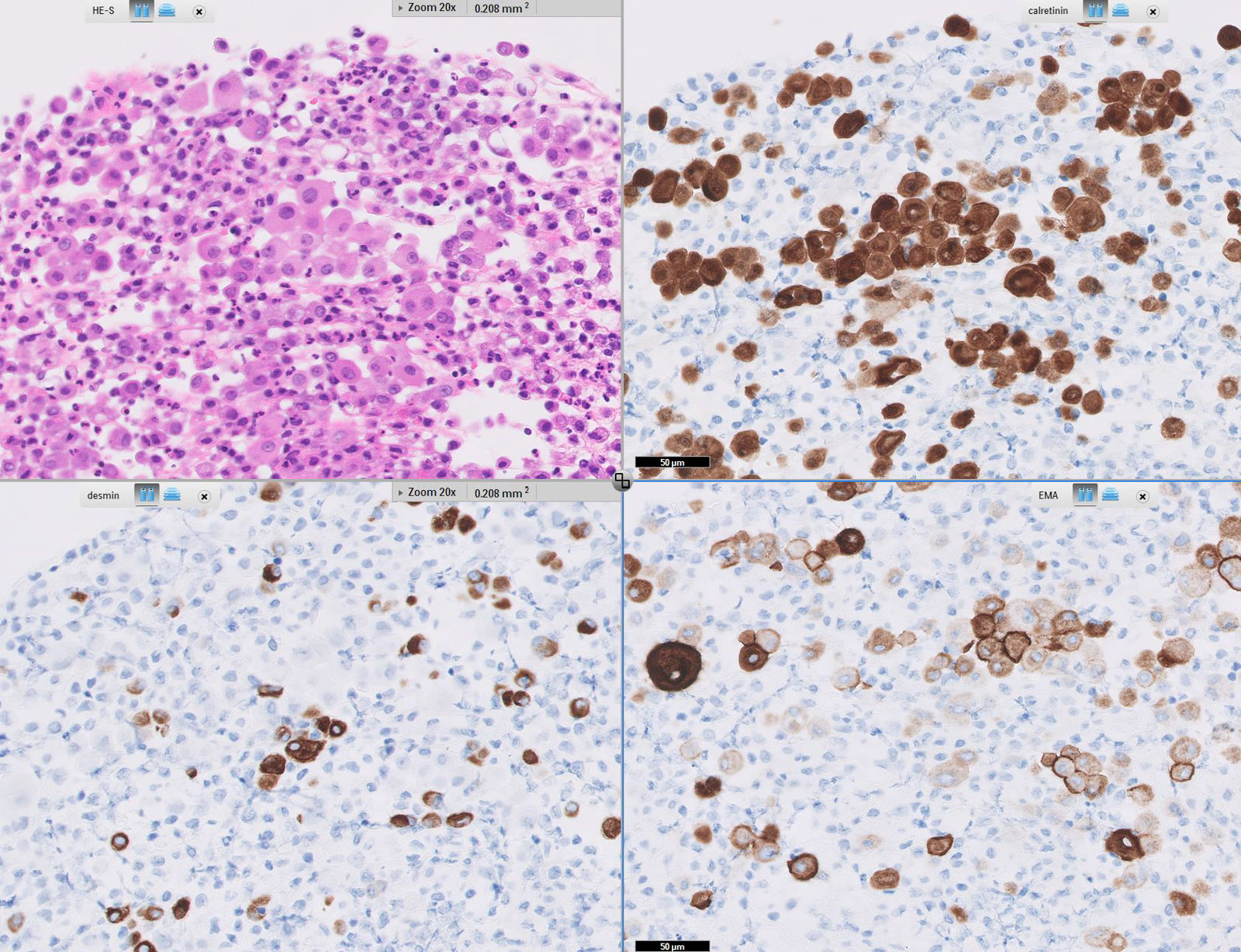
Wt 1 protein is conventionally used as a positive mesothelioma marker. Acute myeloid leukemia cystic partially differentiated nephroblastoma desmoplastic small round cell tumor malignant mesothelioma metanephric adenoma nephrogenic rests ovarian carcinomas serous carcinoma almost all transitional small cell am j surg pathol 2005291034 peritoneal serous carcinoma involving an endometrial polyp 80 am j surg pathol. 3 15 in this case the tumor cells were epithelioid and positive for calretinin wt1 ck56 and d240 staining which confirmed the diagnosis of malignant mesothelioma.
According to their 2012 guidelines mesothelioma tumors test positive for wt1 in 70 to 95 percent of cases. A panel of four markers two positive and two negative selected based upon availability and which ones yield good staining results in a given laboratory is recommended. The antibody reacts with all isoforms of the full length wt1 and also identifies wt1 lacking exon 2 encoded amino acids.
To compare specificity. Wilms tumor 1 protein regulates transcription of other genes and can function both as a transcriptional activator and repressor. The best discriminators among the antibodies considered to be negative markers for mesothelioma are cea moc 31 ber ep4 bg 8 and b723.
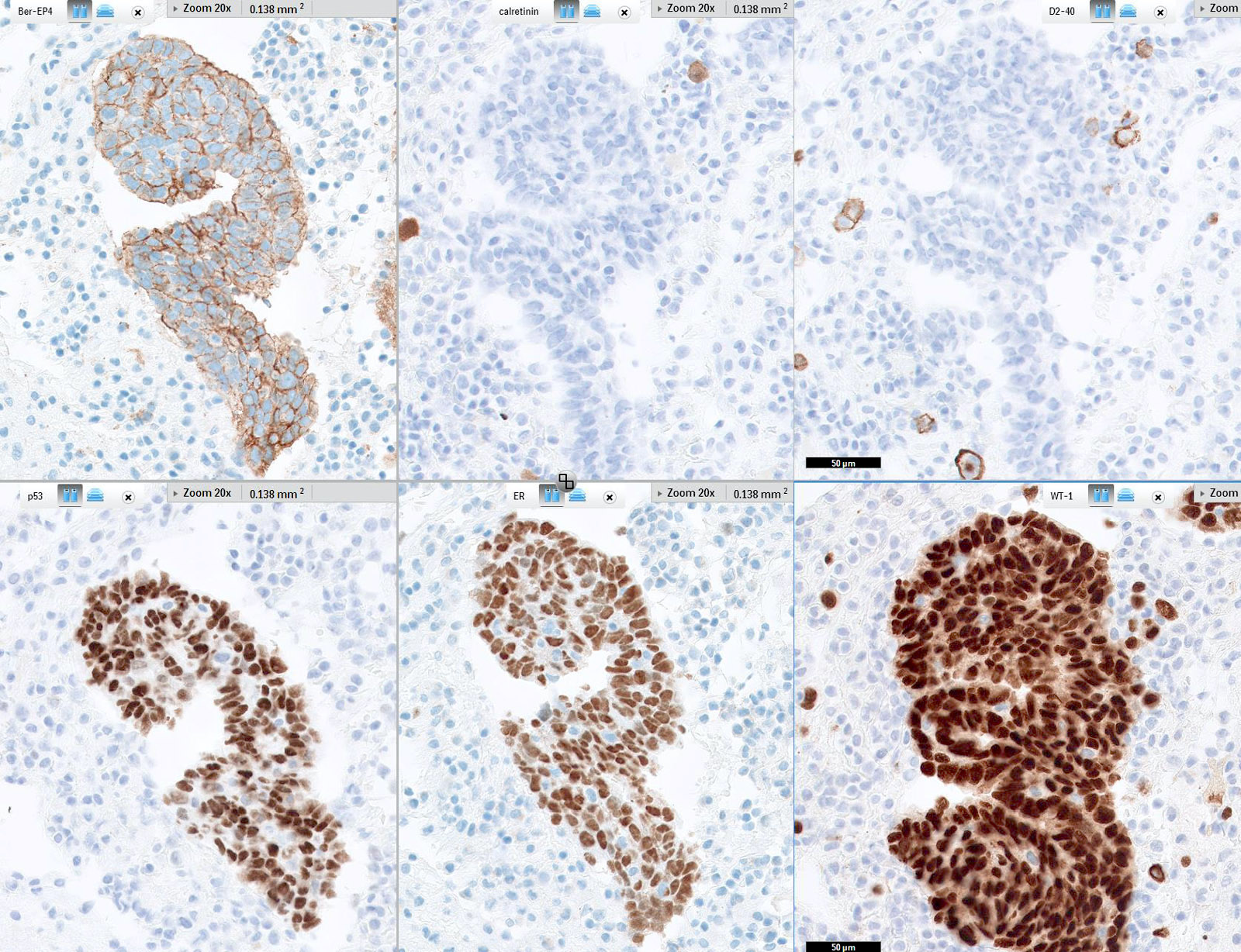
To distinguish malignant mesothelioma adenocarcinoma and reactive benign mesothelium with cytological and histological methods including immunocytochemistry is a major diagnostic challenge. Hbme 1 moc 31 wt1 and calretinin. Nuclear staining for wt1 is highly specific for mesothelioma and in the appropriate clinical setting can be a helpful adjunct in the distinction between adenocarcinomas and mesotheliomas.
Calretinin ck56 wt1 and d240 are considered to be the best indicators for differentiating mesothelioma. The degree of positivity with calretinin may be dependent on the specific antibody utilized. An assessment of recently described markers for mesothelioma and adenocarcinoma.
More From Marple Rubin Family Law
- Cute Monster Colouring Pages
- Coloring Pages Lol Surprise
- Chandler Law
- Free Wiccan Coloring Pages
- Fortnite Coloring Page
Incoming Search Terms:
- Wt1 Antibody Wt1 1434r Nbp2 53182 Novus Biologicals Fortnite Coloring Page,
- First Case Report Of Malignant Peritoneal Mesothelioma And Oral Verrucous Carcinoma In A Patient With A Germline Pten Mutation A Combination Of Extremely Rare Diseases With Probable Further Implications Bmc Medical Fortnite Coloring Page,
- Positive Nuclear Bap1 Immunostaining Helps Differentiate Non Small Cell Lung Carcinomas From Malignant Mesothelioma Abstract Europe Pmc Fortnite Coloring Page,
- Wilms Tumor 1 Susceptibility Wt1 Gene Products Are Selectively Expressed In Malignant Mesothelioma Abstract Europe Pmc Fortnite Coloring Page,
- Anti Wilms Tumor 1 Antibody Wt1 857 Wt1 Fortnite Coloring Page,
- Malignant Mesothelioma A Histomorphological And Immunohistochemical Study Of 24 Cases From A Tertiary Care Hospital In Southern India Hui M Uppin Sg Bhaskar K Kumar Nn Paramjyothi Gk Indian J Cancer Fortnite Coloring Page,


