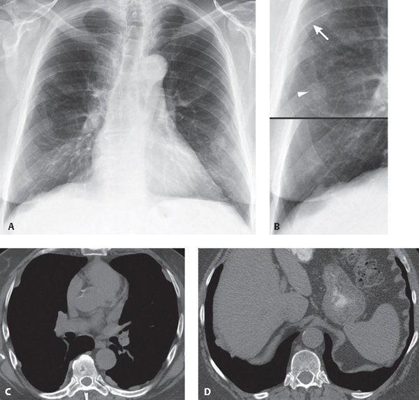
What Is Calcified Pleural Plaques, Left Pleural Effusion And Calcified Pleural Plaques Visible On Chest Download Scientific Diagram
What is calcified pleural plaques Indeed recently has been sought by consumers around us, perhaps one of you personally. People now are accustomed to using the internet in gadgets to view image and video information for inspiration, and according to the title of this post I will talk about about What Is Calcified Pleural Plaques.
- Calcified Pleural Plaques Asbestos Exposure Radiology Case Radiopaedia Org
- Unilateral Pleural Calcifications Are Usually Due To Infection Tb Empyema Hemorrhagic Clinical
- Memory Of World War Ii With Loud Atypical Friction Rub Due To Pulmonary Asbestosis Bmj Case Reports
- Can I Get Compensation For Pleural Plaques National Asbestos Helpline
- Calcified Pleural Plaques On Chest X Ray Youtube
- Bilateral Diffuse Pleural Plaque With Calcifications In Pleural Tuberculosis Tie Mediastinum
Find, Read, And Discover What Is Calcified Pleural Plaques, Such Us:
- Pdf Pleural Plaque Related To Asbestos Mining In Taiwan
- Pleural Plaques Ct
- Close Up Of Calcified Pleural Asbestos Plaques On Chest Xray Stock Photo Download Image Now Istock
- Diseases Of The Chest Wall Pleura And Diaphragm Springerlink
- Pleural Plaques Pulmonology Weebly Com
- Printable Coloring Sheets Pictures To Print And Colour
- Easy Hedgehog Coloring Page
- Most Searched Words For Mesothelioma
- Mark Baker Lawyer
- Dove Coloring Page Printable
If you re looking for Dove Coloring Page Printable you've arrived at the right place. We have 100 images about dove coloring page printable adding pictures, pictures, photos, backgrounds, and much more. In such web page, we also have number of graphics out there. Such as png, jpg, animated gifs, pic art, logo, black and white, transparent, etc.
In 95 to 100 of cases a ct scan can correctly identify patients who have.

Dove coloring page printable. Infection involving the pleura. It is very common for areas of this membrane to become thickened and to accumulate a chalky material if one has been exposed to asbestos. In general doctors tend to prefer this type of imaging test as it is much more sensitive and specific than a chest x ray meaning it can detect.
Abstract pleural thickening has a variety of causes and often must be distinguished from pleural masses while pleural calcifications are frequently the result of chronic infections including bacterial or tuberculous empyema. The pleura is a two layered membrane surrounding the lungs and lining the inside of the rib cage. Pyothoraxempyema tuberculous pleuritis 3.
The pleural plaques of asbestos may be localized soft tissue but frequently calcify with a characteristic radiologic appearance on both chest x ray and ct. Pleural plaques may be calcified or noncalcified but only calcified plaques are readily seen on chest x ray. However pleural plaques usually cause no measurable impairment of pulmonary function or of an.
Pleural calcification can be the result of a wide range of pathology and can be mimicked by a number of conditionsartifacts. Diagnosis with ct scan. The calcification makes pleural plaques very apparent on x rays and they appear radiologically generally at least 10 years after first exposure to asbestos with the extensiveness of plaquing often related to duration and intensity of exposure.
The impacted areas of the lung can be even clearer if the patient has calcified pleural plaques. If you have been exposed to asbestos it is very common for areas of this membrane to become thickened and to accumulate a chalky material. The area where a pleural plaque is located therefore cannot function as normal lung tissue does because it has hardened and can no longer expand as the lungs inflate during breathing.
For pleural plaques this is usually 20 30 years after exposure. A ct scan is the preferred method for diagnosing this condition as it can identify plaques anywhere in the chest even if they are not calcified. If you have pleural plaques it doesn.
These areas are called pleural plaques. Eventually the plaque may become calcified which means calcium salts have built up in the tissue causing it to harden. Calcified pleural plaques appear as translucent white deposits on the lungs in x ray imaging scans.
A ct scan will be able to create a clearer image of the pleural plaques and their extent on the lung. Noncalcified plaque may not be visible at all unless it is very thick. Having the disease does not mean that you are certain to go on to develop a separate more serious asbestos related disease such as mesothelioma or asbestosis.
The pleura is a two layered membrane surrounding your lungs and lining the inside of your rib cage. Typically with sparing of the costophrenic angles. Like other asbestos related diseases pleural plaques does not develop until decades after initial exposure.
More From Dove Coloring Page Printable
- Cooking Coloring Pages
- Child Support And Custody Lawyer
- Kim Kardashian Lawyer
- Unicorn Coloring Sheets
- Pleural Mesothelioma Story
Incoming Search Terms:
- Can I Get Compensation For Pleural Plaques National Asbestos Helpline Pleural Mesothelioma Story,
- Investigating Pleural Thickening The Bmj Pleural Mesothelioma Story,
- Calcified Pleural Plaques Radiology Case Radiopaedia Org Pleural Mesothelioma Story,
- Asbestos When The Dust Settles An Imaging Review Of Asbestos Related Disease Radiographics Pleural Mesothelioma Story,
- Calcified Pleural Plaques On Chest X Ray X Rays Case Studies Ctisus Ct Scanning Pleural Mesothelioma Story,
- Radiology Corner European Respiratory Society Pleural Mesothelioma Story,


