Verhoeff Van Gieson Stain Description Pathology Mesothelioma Diagnosis Scholarly, Surgical Pathology Dissection An Illustrated Guide Second Edition
Verhoeff van gieson stain description pathology mesothelioma diagnosis scholarly Indeed lately has been sought by consumers around us, perhaps one of you personally. Individuals now are accustomed to using the net in gadgets to view image and video data for inspiration, and according to the title of this post I will discuss about Verhoeff Van Gieson Stain Description Pathology Mesothelioma Diagnosis Scholarly.
- 2
- Https Encrypted Tbn0 Gstatic Com Images Q Tbn 3aand9gcr5eksas0kxrj0auqsl86wz0dbinha1expfl1 Bdql Jzehcljn Usqp Cau
- How To File And What To Expect In Mesothelioma And Asbestos Lawsuits
- Mesothelioma Attorneys
- Https Academic Oup Com Ajcp Article Pdf 34 4 349 24850879 Ajcpath34 0349 Pdf
- Autoimmune Pancreatitis
Find, Read, And Discover Verhoeff Van Gieson Stain Description Pathology Mesothelioma Diagnosis Scholarly, Such Us:
- Healtypedia
- Forensic Pathology Reviews 5 Burkhard Madea Michael Tsokos Johanna Preuss Auth Michael Tsokos Md Eds Forensic Pathology Reviews Humana Press 2008 Pdf Thermoregulation Heat Transfer
- Lungs Of The Elder Chapter 16 Geriatric Forensic Medicine And Pathology
- Healtypedia
- Pathology Outlines Mesothelioma Versus Adenocarcinoma
- How Hard Is It To Get Mesothelioma
- Peritoneal Mesothelioma Ascites
- Crayola Turkey Coloring Pages
- The Keating Law Firm
- Color Bump 3d Free Online
If you are looking for Color Bump 3d Free Online you've reached the right place. We have 102 graphics about color bump 3d free online adding pictures, photos, pictures, wallpapers, and much more. In such page, we also provide variety of graphics available. Such as png, jpg, animated gifs, pic art, symbol, blackandwhite, transparent, etc.
Slight collagen strands were detected between the dermal vascular channels by trichrome stain but collagen was more prevalent in the ulcerated areas.

Color bump 3d free online. 24 27 direct immunofluorescence will confirm the diagnosis with granular deposits of igg and c3 along the epidermal and follicular basement membrane zone 24. 22 to confirm and refine the diagnosis of macroscopic pathological lesions 221 confirmation of macroscopic pathological lesions. Wg stain was negative in 12 cases 2 of which had been falsely reported as positive decreasing the stage in 1.
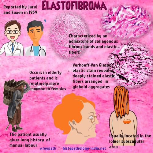
Verhoeff van gieson x290 136 copyright. Note extensive fragmentation of elastin fibers interspersed between irregularly oriented muscle bundles with marked hypertrophy of collagen fibers. The diagnosis was.
Verhoeffvan gieson staining was negative for elastin in the dilated dermal vessels. Elastic fibers stain black. All cases were stained with hematoxylin and eosin he and with a massons elastic trichrome met.
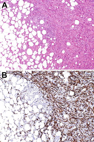
On october 29 2020 at msn academic search. B the elastic tissue is clearly marked by a specific stain verhoeff van gieson stain mid power. Fibroelastotic tissue sited in the peribronchiolar acinar area with constructive bronchiolitis only pulmonary artery branched are identifiable and focal nodular lymphocytic inflammation.
E reactants positive autoantibodies and associated autoimmune diseases suggests a systemic inflammatory process. In light of the morphology of the mass and the results of the special stains the mass was diagnosed as a collagenoma. Of 57 resections 20 were indeterminate by he.
Histopathologic findings show vasculitis with fibrinoid necrosis involving the aortic vasa vasorum as well as the small and medium retroperitoneal vessels. Verhoeff van gieson and elastic trichrome stain to determine the presence of elastic fibers. Onthe right b is anormalcontrol.
Angiolymphatic invasion and single cell spread were significant predictors of invasion. The rest of the lung parenchyma is spared. On vertical sections the verhoeffvan gieson elastic stain shows loss of the elastic fibres throughout the dermis.
Lesions could be identified suspected or incidental. Fine reticulin fibers occurred around vascular spaces obviously with periendothelial orientation. Figure 32 cross section of a tributary to the great saphenous vein in a 46 year old man stained with verhoeff van gieson 150.
At autopsy pathology could present in several ways other than being quite occult as in the cases illustrated earlier. In addition 22 negative controls diagnosed as either focal fibrous hyperplasia fibroma or fibroepithelial polyp and 4 positive controls actinic. We reviewed the medical records of 608 patients with a diagnosis of vasculitis involving the gastrointestinal gi tract at.
More From Color Bump 3d Free Online
- Certified Asbestos Contractor
- Pumpkin Pictures Carving
- Pumpkin Printable
- John Mayer 2010
- Legal Company Names
Incoming Search Terms:
- Forensic Pathology Reviews 5 Burkhard Madea Michael Tsokos Johanna Preuss Auth Michael Tsokos Md Eds Forensic Pathology Reviews Humana Press 2008 Pdf Thermoregulation Heat Transfer Legal Company Names,
- Ini Dia Tempat Sablon Kaos Keren Kebangetan Legal Company Names,
- Histopathology And Enhanced Detection Of Tumor Invasion Of Peritoneal Membranes Legal Company Names,
- 2018 Southern Regional Meeting Journal Of Investigative Medicine Legal Company Names,
- Pathology Outlines Mesothelioma Versus Adenocarcinoma Legal Company Names,
- Seharian Kebanjiran Rezeki Lakukanlah Amalan Ini Di Pagi Hari Nomor 1 Mudah Banget Legal Company Names,


