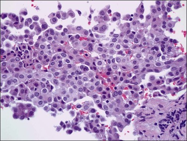
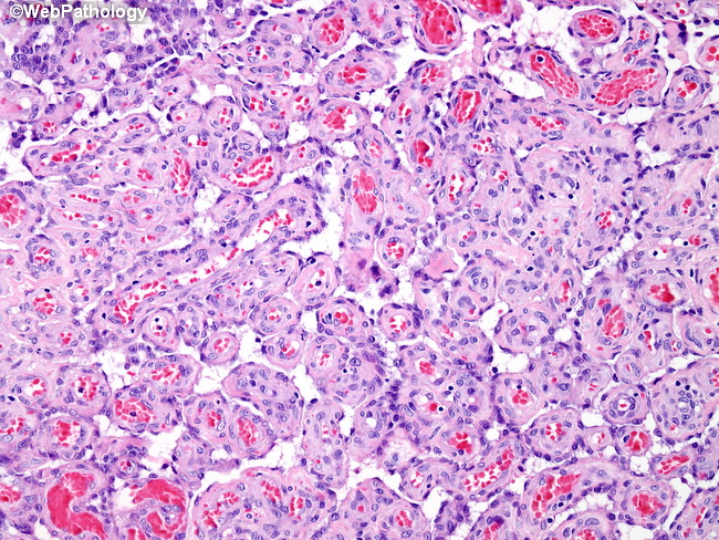
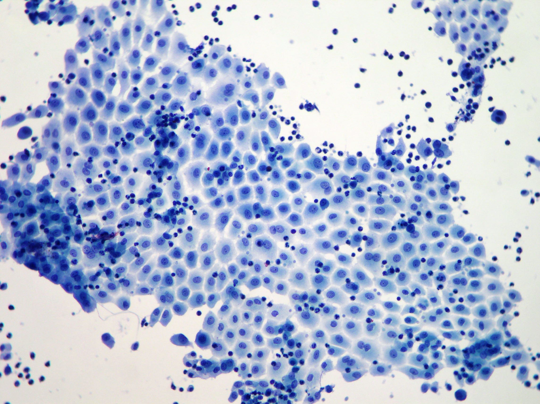
Reactive Mesothelioma Cells, Sheet Of Reactive Mesothelial Cells Note Owindowso Between Cells And Download Scientific Diagram
Reactive mesothelioma cells Indeed recently is being hunted by consumers around us, perhaps one of you. Individuals now are accustomed to using the net in gadgets to see video and image data for inspiration, and according to the name of the article I will talk about about Reactive Mesothelioma Cells.
- Pb Reactive Mesothelial Hyperplasia Mimicking Mesothelioma In An African Green Monkey Chlorocebus Aethiops
- Http Handouts Uscap Org 2016 Cm06 Daci 1 Pdf
- Jcdr Adenocarcinoma Immunocytochemistry Reactive Mesothelial Cells Serous Effusions
- File Reactive Mesothelial Cells High Mag Jpg Wikimedia Commons
- Reliability Of P 16 Calretinin And Claudin 4 Immunocytochemistry In Diagnostic Verification Of Effusion Cytology
- Differentiation Of Mesothelial Cells Into Macrophage Phagocytic Cells In A Patient With Clinical Sepsis
Find, Read, And Discover Reactive Mesothelioma Cells, Such Us:
- The Cell Morphological Characteristics Of Adenocarcinoma Cells Download Scientific Diagram
- Reactive Mesothelial Cells With Atypia Mesothelial Cells May Contain Multiple Nuclei Which May Cause Concern For Mal Hematology Medical Laboratory Immunology
- Https Www Rcpath Org Asset Ed8cdd8d 8d04 4b82 Ad48d585e2f023be
- Differentiation Of Mesothelial Cells Into Macrophage Phagocytic Cells In A Patient With Clinical Sepsis
- Body Cavityfluids Chapter 3 Differential Diagnosis In Cytopathology
- Law Office Artwork
- Princess Pumpkin Ideas
- Ellen Brockovich Film
- Loculated Pleural Effusion Mesothelioma
- Lebanon Mesothelioma Lawyer
If you are looking for Lebanon Mesothelioma Lawyer you've come to the perfect location. We ve got 104 graphics about lebanon mesothelioma lawyer including pictures, photos, pictures, wallpapers, and much more. In these web page, we also provide variety of images available. Such as png, jpg, animated gifs, pic art, symbol, black and white, transparent, etc.

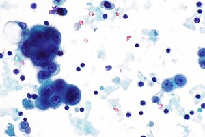
Pleural Fluid Clusters Of Atypical Reactive Mesothelial Cells From A Download Scientific Diagram Lebanon Mesothelioma Lawyer
A mm epithelioid type he 40x objective.
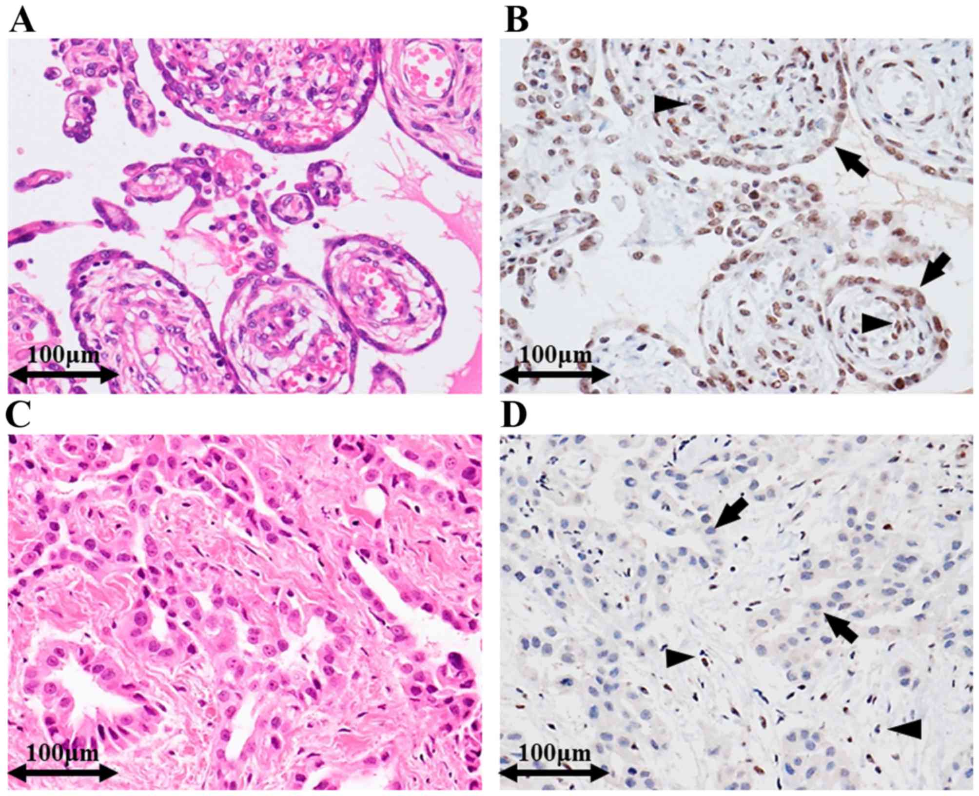
Lebanon mesothelioma lawyer. E rmcs in peritoneal. When these surfaces become irritated or injured mesothelial cells can proliferate and take on a variety of morphologic and cytologic appearances. Of 217 cases circulated among all members of the uscanadian mesothelioma reference panel there was some disagreement about whether the process was benign or malignant in 22 of cases.
The nuclear features are helpful if the mesothelioma is poorly differentiated. Archival paraffin embedded cell blocks of pleural and peritoneal fluids from 52 patients with malignant mesothelioma mm and 64 patients with reactive mesothelial hyperplasia mh were retrieved. In biopsy tissue discrimination between reactive mesothelial hyperplasia and epithelial mesothelioma can pose a major problem for the surgical pathologist.
Confidence in the diagnosis is often proportional to the amount of tissue available for study and depends largely on findings of invasion and t. 1 frank invasion is regarded as the most. Ihc stains included desmin epithelial membrane antigen ema glucose transport protein 1 glut 1 ki67 and p53.
Reactive mesothelial cells can be found when there is an infection or an inflammatory response present in a body cavity. D presence of nuclear bap1 ihc staining in mm. Intracytoplasmic vacuole cells were classified as.
This condition can be due to the presence of a bacterial viral or fungal infection. In situ hybridisation against p16 is a promising method of differentiating malignant mesothelioma from reactive mesothelium. Psammomatous calcifications can be seen in both conditions.
B absence of nuclear bap1 ihc staining for in malignant cells. The distinction between reactive mesothelial hyperplasia mh and malignant mesothelioma mm may be very difficult based only on histologic and morphologic findings. Mesothelial cells are mesodermally derived epithelial cells that line body cavities pleura pericardium and peritoneum.
Note internal control of rmcs and histiocytes. Fibrous pleurisy may show spaces mimicking fat fake fat surrounded by cytokeratin positive spindle cells a source of confusion with desmoplastic mesothelioma. C mm papillary type.
Under normal conditions mesothelial cells form a flat single uniform layer. Reactive mesothelial cells can closely mimic both mesothelioma and carcinoma on histopathological and cytological examination of tissues and effusions respectively and it is widely recognised in human and veterinary medicine that distinguishing reactive from neoplastic mesothelium can be very challenging. However in well differentiated cases malignant mesothelioma may show features identical to benign and reactive mesothelial cells.
Mesothelioma mm and reactive mesothelial cell rmc proliferations in cytology samples.
More From Lebanon Mesothelioma Lawyer
- Which Type Of Asbestos Causes Mesothelioma
- Church Coloring Pages Free
- Food Coloring Sheets
- Gad3 Mesothelioma
- Simple Feather Coloring Page
Incoming Search Terms:
- Reactive Mesothelial Cells In Pleural Effusion Showing Variation In Download Scientific Diagram Simple Feather Coloring Page,
- Http Jtd Amegroups Com Article Viewfile 25454 Pdf Simple Feather Coloring Page,
- Http Www Asl5 Liguria It Portals 0 Anatomiapatologica2015 20150924 Effusion Cytology Pdf Simple Feather Coloring Page,
- Home Simple Feather Coloring Page,
- The Panorama Of Different Faces Of Mesothelial Cells Basicmedical Key Simple Feather Coloring Page,
- Https Encrypted Tbn0 Gstatic Com Images Q Tbn 3aand9gcrgrvn 3be6wylpxdte0su7bvf9lduonoausevalghb5uds Xu6 Usqp Cau Simple Feather Coloring Page,
