Reactive Mesothelial Cells Vs Mesothelioma Ihc, Https Patologi Com Guideline 20mesotheliom Pdf
Reactive mesothelial cells vs mesothelioma ihc Indeed lately is being sought by users around us, maybe one of you. People are now accustomed to using the net in gadgets to view image and video information for inspiration, and according to the title of the article I will talk about about Reactive Mesothelial Cells Vs Mesothelioma Ihc.
- What S New In Mesothelioma Pathologica Journal Of The Italian Society Of Anatomic Pathology And Diagnostic Cytopathology
- The Diagnostic Utility Of Immunohistochemistry In Distinguishing Between Epithelioid Mesotheliomas And Squamous Carcinomas Of The Lung A Comparative Study Modern Pathology
- Https Www Hkiap Org Wp Content Uploads Lecture Notes 2019 20special 20cytology 20workshop Mesothelioma 20how 20far Pdf
- Utility Of Survivin Bap1 And Ki 67 Immunohistochemistry In Distinguishing Epithelioid Mesothelioma From Reactive Mesothelial Hyperplasia
- Mesothelial Hyperplasia An Overview Sciencedirect Topics
- Https Www Surgpath Theclinics Com Article S1875 9181 10 00011 5 Pdf
Find, Read, And Discover Reactive Mesothelial Cells Vs Mesothelioma Ihc, Such Us:
- Webpathology Com A Collection Of Surgical Pathology Images
- Immunohistochemical Reaction Of Reactive Mesothelium Mesothelioma Download Table
- Highly Expressed Ezh2 In Combination With Bap1 And Mtap Loss As Detected By Immunohistochemistry Is Useful For Differentiating Malignant Pleural Mesothelioma From Reactive Mesothelial Hyperplasia Lung Cancer
- Non Neoplastic Reactive Mesothelial Cells Dog With Non Neoplastic Download Scientific Diagram
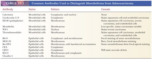
- Http Phenopath Com Uploads Pdf Mesothelioma Vs Adenocarcinoma Pdf
- Mesothelioma Vs Non Small Cell Lung Cancer
- Most Common Cause Of Mesothelioma
- Cinderella Coloring Sheet
- Popular Halloween Costumes 2019
- Hartford Mesothelioma Claim
If you re looking for Hartford Mesothelioma Claim you've reached the ideal location. We have 104 images about hartford mesothelioma claim including images, photos, pictures, backgrounds, and more. In such page, we also have variety of graphics out there. Such as png, jpg, animated gifs, pic art, logo, black and white, translucent, etc.
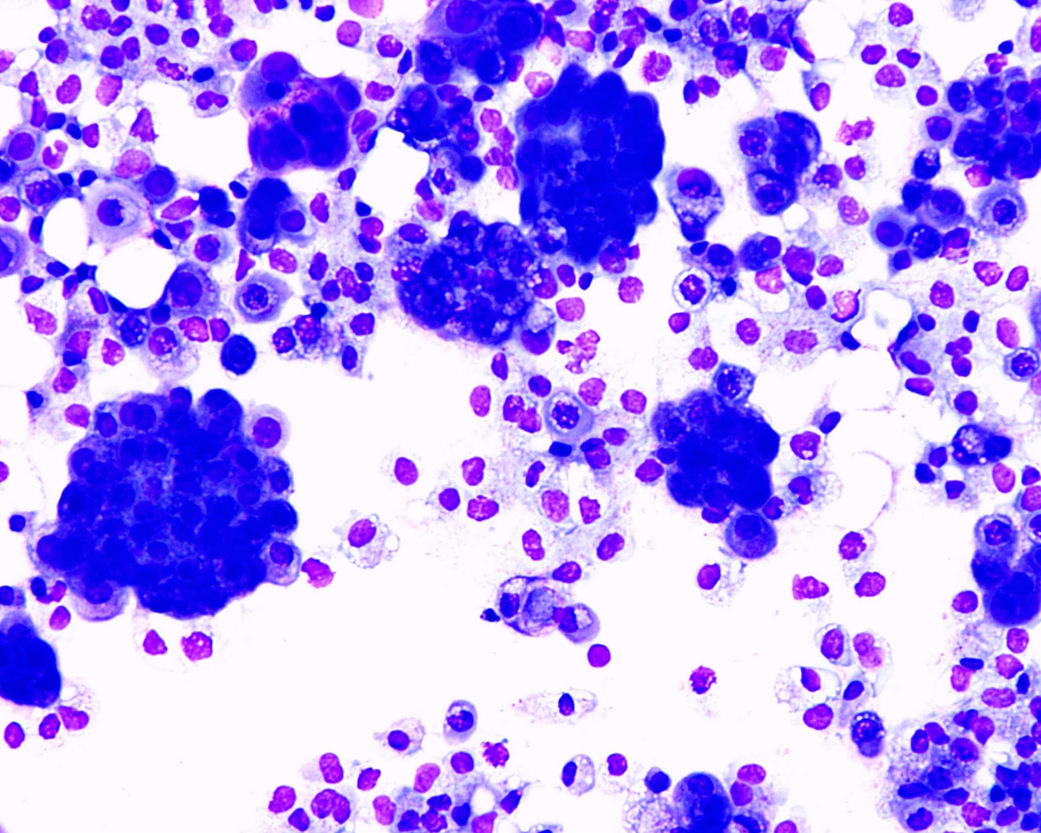
3 d clusters of cells strongly.

Hartford mesothelioma claim. 163299 305 26 kato y tsuta k seki k et al. The distinction of benign from malignant mesothelial proliferations in cytologic specimens can be. 1 frank invasion is regarded as the most.
Lin md phd. Cytology only diagnosis biopsies immunohistochemistry discrimination between mesothelioma and reactive mesothelial hyperplasia and biomarkers j clin pathol. Archival paraffin embedded cell blocks of pleural and peritoneal fluids from 52 patients with malignant mesothelioma mm and 64 patients with reactive mesothelial hyperplasia mh were retrieved.
The differential diagnosis of epithelial type mesothelioma from adenocarcinoma and reactive mesothelial proliferation. Focal hyperchromasia is seen in reactive mesothelial cells. The aims of this study were to clarify the usefulness of immunohistochemistry in the differential diagnosis of epithelioid mesothelioma with a solid growth pattern solid epithelioid mesothelioma sem and poorly differentiated squamous cell carcinoma pdscc and to confirm the validity of a specific type of antibody panel.
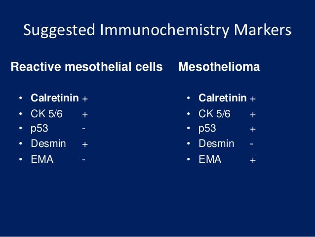
Ihc stains included desmin epithelial membrane antigen ema glucose transport protein 1 glut 1 ki67 and p53. Additionally we aimed to clarify the pitfalls of. The distinction between reactive mesothelial hyperplasia mh and malignant mesothelioma mm may be very difficult based only on histologic and morphologic findings.
The use of immunohistochemistry to distinguish reactive mesothelial cells from malignant mesothelioma in cytologic effusions farnaz hasteh md 1. Large nc ratios may be seen in reactive mesothelial cells. Immunohistochemical detection of glut 1 can discriminate between reactive mesothelium and malignant mesothelioma.
Of 217 cases circulated among all members of the uscanadian mesothelioma reference panel there was some disagreement about whether the process was benign or malignant in 22 of cases. The morphological evaluation of cytological specimens from body cavity fluids presents difficulties in the differential diagnosis between benign reactive mesothelial rm cells and adenocarcinoma ac or malignant mesothelioma mm. Focal macronucleoli are seen in reactive mesothelial cells.
The aim of our study was to investigate whether a panel of five dif. Nc ratio may be normal in mesothelioma.

Reliability Of P 16 Calretinin And Claudin 4 Immunocytochemistry In Diagnostic Verification Of Effusion Cytology Hartford Mesothelioma Claim
More From Hartford Mesothelioma Claim
- Telephone Coloring Pages
- How Long Of Exposure To Asbestos Is Dangerous
- Surviving Mesothelioma And Other Cancers A Patients Guide Pdf
- Drawing Forky Coloring Page
- Mesothelioma Prognosis Stage 4
Incoming Search Terms:
- Http Phenopath Com Uploads Pdf Mesothelioma Vs Adenocarcinoma Pdf Mesothelioma Prognosis Stage 4,
- 2 Mesothelioma Prognosis Stage 4,
- Mesothelial Hyperplasia An Overview Sciencedirect Topics Mesothelioma Prognosis Stage 4,
- Use Of Panel Of Markers In Serous Effusion To Distinguish Reactive Mesothelial Cells From Adenocarcinoma Subbarayan D Bhattacharya J Rani P Khuraijam B Jain S J Cytol Mesothelioma Prognosis Stage 4,
- Https Www Hkiap Org Wp Content Uploads Lecture Notes 2019 20special 20cytology 20workshop Mesothelioma 20how 20far Pdf Mesothelioma Prognosis Stage 4,
- Mesothelial Cytopathology Libre Pathology Mesothelioma Prognosis Stage 4,


