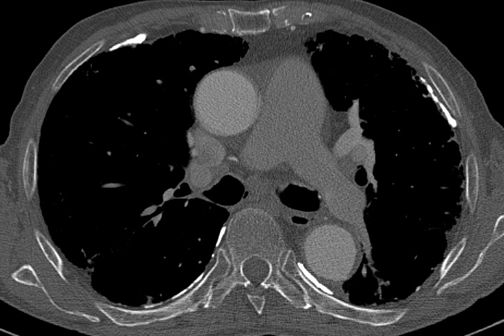
Pleural Plaques Nhs, Nhs To Offer Mesothelioma Patients Alternative Immunotherapy During Covid Pandemic Fieldfisher
Pleural plaques nhs Indeed recently has been sought by consumers around us, maybe one of you personally. People now are accustomed to using the net in gadgets to view video and image information for inspiration, and according to the title of the post I will talk about about Pleural Plaques Nhs.
- Nhs To Offer Mesothelioma Patients Alternative Immunotherapy During Covid Pandemic Fieldfisher
- Pictorial Essay Of Radiological Features Of Benign Intrathoracic Masses
- Non Malignant Asbestos Related Diseases A Clinical View Rcp Journals
- Epos Trade
- Non Malignant Asbestos Related Diseases A Clinical View Rcp Journals
- Postero Anterior And Lateral Chest Radiographs Demonstrating Extensive Download Scientific Diagram
Find, Read, And Discover Pleural Plaques Nhs, Such Us:
- Find Out About Symptoms Diagnosis And Treatments For Mesothelioma Action On Asbestos Industrial Injury Disease
- Http Pdf Posterng Netkey At Download Index Php Module Get Pdf By Id Poster Id 108078
- Asbestosis
- Find Out About Symptoms Diagnosis And Treatments For Mesothelioma Treatment Of Mesothelioma Action On Asbestos Industrial Injury Disease
- High Resolution Ct Shows Minimal Pleural Plaque Arrow Overlying A Download Scientific Diagram
- Cardiac Mesothelioma
- Class Action Mesothelioma
- Patent Lawyer Nyc
- Metastatic Mesothelioma Ct Chest
- Disney Colouring Pages To Print
If you re looking for Disney Colouring Pages To Print you've arrived at the ideal location. We have 100 graphics about disney colouring pages to print adding pictures, photos, photographs, backgrounds, and more. In such webpage, we also provide variety of images available. Such as png, jpg, animated gifs, pic art, logo, black and white, translucent, etc.

The Calcified Lung Nodule What Does It Mean Topic Of Research Paper In Clinical Medicine Download Scholarly Article Pdf And Read For Free On Cyberleninka Open Science Hub Disney Colouring Pages To Print
If pleural effusion does not clear up as your pleurisy is treated or youre very short of breath the fluid may need to be drained by inserting a needle or tube through the.

Disney colouring pages to print. One layer lines the inside of your chest wall. The inner layer the visceral pleura covers the lung and the outer layer the parietal pleura lines the ribcage and diaphragm. They very rarely cause.
It is important to note that although pleural plaques denote that an exposure to asbestos they are also asymptomatic which means they do not usually have any symptoms at all and that as of themselves they are also harmless with thousands of people already having. The pleura is a thin membrane inside the ribcage surrounding each lung. Pleural plaques are typically identified through imaging scans.
The two layers are normally in contact and secrete a. Pleural plaques are areas of scarring or calcification on the pleura. In some patients the latency period is less than 10 years.
Pleural plaques can be a worrying condition for sufferers as they signify that a person has been exposed to asbestos in the past. These areas are called pleural plaques. Pleural plaques are areas of hyalinized collagen fibers and form in the pleura.
Pleural effusion can lead to shortness of breath that gets progressively worse. Commonly the condition may first be identified in a chest radiograph or x ray that shows thickened areas of the lung with concrete edges which some researchers have noted can look a bit like a holly leaf. The pleura is a two layered membrane surrounding your lungs and lining the inside of your rib cage.
The plaques themselves are harmless they do not cause any symptoms. Between the two layers of pleura your pleural cavity is a tiny amount of fluid. Specifically pleural plaques are described as localized fibrous deposits that thicken the lung lining.
It is most unusual for pleural plaques to cause any symptoms or explain any clinical signs. However in most cases they mean that you have been exposed to asbestos either in your work or occasionally in your home at some time in the past. This acts like lubricating oil between your lungs and your chest wall as they move when you breathe.
Pleural plaques refer to the presence of scar tissue on the parietal pleura. Imaging scans may show pleural plaques 20 to 30 years after long term inhalation of asbestos fibers. This is more likely if pleurisy is caused by pulmonary embolism or a bacterial infection.
It consists of two layers. If you have been exposed to asbestos it is very common for areas of this membrane to become thickened and to accumulate a chalky material. They develop as white lesions with a rubbery consistency though may become calcified or hardened over more time.
The pleura is a thin membrane with two layers. They are the commonest manifestation of past exposure to asbestos and are often reported as an incidental finding on a chest x ray which has been arranged for some other reason. Pleural plaques are areas of thickening of the lining between the lung and chest wall.
They usually develop on the parietal pleura that is the layer of pleura lining the chest cavity. They are the most common sign of asbestos exposure.
More From Disney Colouring Pages To Print
- Fortnite Drift Costume
- Gladney Beautyrest Black Hybrid
- What To Do If You Have Breathed In Asbestos
- Fruit Coloring
- Indolent Mesothelioma
Incoming Search Terms:
- Https Www Resmedjournal Com Article S0954 6111 17 30033 1 Pdf Indolent Mesothelioma,
- Http Pdf Posterng Netkey At Download Index Php Module Get Pdf By Id Poster Id 108078 Indolent Mesothelioma,
- Https Www Resmedjournal Com Article S0954 6111 17 30033 1 Pdf Indolent Mesothelioma,
- Memory Of World War Ii With Loud Atypical Friction Rub Due To Pulmonary Asbestosis Bmj Case Reports Indolent Mesothelioma,
- Out In The Cold Lung Disease The Hidden Driver Of Nhs Winter Pressure British Lung Foundation Indolent Mesothelioma,
- Syndrome Of Pleural And Retrosternal Bridging Fibrosis And Retroperitoneal Fibrosis In Patients With Asbestos Exposure Thorax Indolent Mesothelioma,


