Pericardial Mesothelioma On X Ray, Types Of Mesothelioma Koonz Mckenney Johnson Depaolis Llp
Pericardial mesothelioma on x ray Indeed lately is being hunted by consumers around us, maybe one of you personally. People are now accustomed to using the internet in gadgets to see image and video information for inspiration, and according to the title of the post I will talk about about Pericardial Mesothelioma On X Ray.
- Malignant Pleural Mesothelioma In A Child Sciencedirect
- Visual Guide To Malignant Mesothelioma
- Malignant Pleural Effusion Pulmonology Advisor
- Https Www Resmedjournal Com Article S0954 6111 17 30033 1 Pdf
- Https Www Karger Com Article Pdf 228894
- Mesothelioma Radiology Reference Article Radiopaedia Org
Find, Read, And Discover Pericardial Mesothelioma On X Ray, Such Us:
- How Mesothelioma And Lung Cancer Hide From Early Detection
- Mesothelioma Radiology Reference Article Radiopaedia Org
- Mesothelioma Mesothelioma Radiology Pulmonology
- Malignant Mesothelioma Pericardium
- Hemothorax Wikipedia
- Mathur Law Firm
- Michael Goldberg Attorney
- Endothelium Vs Mesothelium
- Mesothelioma Pulmonary Fibrosis
- Cool Scary Pumpkins
If you are searching for Cool Scary Pumpkins you've come to the perfect place. We have 103 images about cool scary pumpkins including images, photos, pictures, backgrounds, and much more. In such page, we additionally have number of images available. Such as png, jpg, animated gifs, pic art, symbol, blackandwhite, translucent, etc.
Unfortunately many pericardial mesothelioma patients arent diagnosed until an autopsy is performed with one report estimating about 10 20 of cases are properly diagnosed before a patients death.

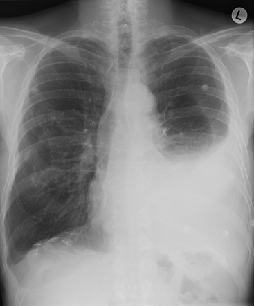
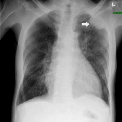
Cool scary pumpkins. Most tumors are easier to spot because an x ray will show a distinct lump or bulge connected to the wall of an organ. Tests of this fluid can determine. Given the presence of the mesothelium in different parts of the body mesothelioma can arise in various locations 17.
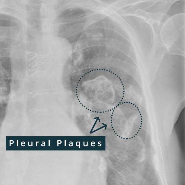
Pleural mesothelioma 90 covered in this article. There are five primary types of imaging tests used during a mesothelioma diagnosis including x rays mri scans ct scans pet scans and ultrasounds. Similarly to peritoneal and pleural mesothelioma diagnosis typically begins with imaging tests like x rays and ct scans.
Given the presence of the mesothelium in different parts of the body mesothelioma can arise in various locations 17. However their results are not detailed enough to diagnosis a mass as mesothelioma pericardial. A chest ct scan or pet scan can.
The particles of the chest x ray are called photons which are absorbed at different rates. The telltale symptoms of pericardial mesothelioma are usually found with visual imaging tests. This is often detected with an echocardiogram x rays or a ct scan of the chest.
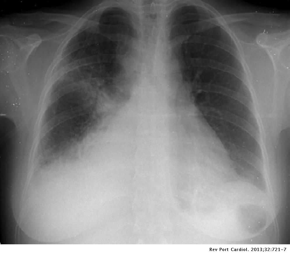
Learn about symptoms prognosis and treatment at maa center. Most tumors arise from the pleura and so this article will focus on pleural mesothelioma. With pericardial mesothelioma however the disease simply makes the heart look larger making it exceptionally hard to provide an accurate diagnosis.
A biopsy of the affected tissue is often used to confirm or rule out the presence of mesothelioma in the lining of the heart. These diagnostic imaging tests are often used to identify tumors and the location of the cancer. Mesothelioma also known as malignant mesothelioma is an aggressive malignant tumor of the mesothelium.
Are usually first noticed in an x ray or ct scan. The next logical step is to conduct imaging tests beginning with an x ray to obtain an understanding of the hearts health. Pleural mesothelioma 90 covered in this article.
Typical symptoms seen in pericardial mesothelioma include. One common sign is pericardial fluid buildup around the heart. Chest x ray flow throughout the body at a frequency much more condensed than light.
After the x ray a ct scan and mri will likely be conducted. X ray ct scan or mri. This procedure uses a needle to remove fluid from the sac around the heart.
Please call us for help at 1 866 371 8506. The final result is a negative of the internal structures of the human body. Imaging scans are non invasive and act as a stepping stone in the diagnostic process for mesothelioma patients.
An x ray provides a flat 2d image of bones and soft tissue.
More From Cool Scary Pumpkins
- Pumpkin Decorating Pictures
- Emoji Pumpkin Painting Ideas
- Gene Therapy For Mesothelioma And Lung Cancer
- Shipping Law Firms
- Tom And Jerry Coloring Pages Online Free
Incoming Search Terms:
- Cardiac Mri Tom And Jerry Coloring Pages Online Free,
- Posterior Anterior And Lateral Chest X Ray With An Enlarged Cardiac Download Scientific Diagram Tom And Jerry Coloring Pages Online Free,
- Diagnosis And Therapeutic Management Of Patients With Cardiac Tamponade And Constrictive Pericarditis Revista Espanola De Cardiologia English Edition Tom And Jerry Coloring Pages Online Free,
- Primary Pericardial Mesothelioma A Rare Entity Tom And Jerry Coloring Pages Online Free,
- How Mesothelioma And Lung Cancer Hide From Early Detection Tom And Jerry Coloring Pages Online Free,
- Pleural Neoplasms Radiology Key Tom And Jerry Coloring Pages Online Free,


