Pathology Stains For Mesothelioma, Webpathology Com A Collection Of Surgical Pathology Images
Pathology stains for mesothelioma Indeed lately has been hunted by users around us, perhaps one of you personally. Individuals are now accustomed to using the internet in gadgets to see video and image information for inspiration, and according to the title of the article I will talk about about Pathology Stains For Mesothelioma.
- Bap1 Immunohistochemistry And P16 Fish Results In Combination Provide Higher Confidence In Malignant Pleural Mesothelioma Diagnosis Roc Analysis Of The Two Tests Hida 2016 Pathology International Wiley Online Library
- Pathology Examination Surviving Mesothelioma
- 2
- Https Www Rcpa Edu Au Events Pathology Update International Guest Speakers Presentations Prof Jeffrey Myers Prof Jeffrey Myers Diagnosis Of Malignant Mesothel Aspx
- Pathology Of Mesothelioma Springerlink
- Utility Of Survivin Bap1 And Ki 67 Immunohistochemistry In Distinguishing Epithelioid Mesothelioma From Reactive Mesothelial Hyperplasia
Find, Read, And Discover Pathology Stains For Mesothelioma, Such Us:
- 2
- Eposters Rare Diagnosis Of Malignant Mesothelioma In A Young Patient With Rheumatoid Arthritis
- Abdominal Tissue Slides Malignant Mesothelioma Nbp2 77445 Novus Biologicals
- Malignant Pleural Mesothelioma Presenting With Cardiac Tamponade A Rare Case Report And Review Of The Literature
- Webpathology Com A Collection Of Surgical Pathology Images
- Mesothelioma Risk If Had Lung Cancer
- Mickey Mouse Colouring Pictures
- Easy Scary Pumpkin Faces To Carve
- Giraffe Coloring Pages Printable
- Cool Coloring Sheets Hard
If you re looking for Cool Coloring Sheets Hard you've reached the perfect place. We have 104 graphics about cool coloring sheets hard adding pictures, photos, photographs, backgrounds, and more. In such page, we also provide variety of graphics out there. Such as png, jpg, animated gifs, pic art, logo, black and white, translucent, etc.
The international mesothelioma interest group imig states a definitive diagnosis of asbestos cancer must include immunohistochemical testing.

Cool coloring sheets hard. This scientific practice also reveals which cell types are present in the tissue or fluid samples. Mesothelioma pathology provides a full picture of the cancer contributing to a more accurate diagnosis and an informed treatment plan. This stain will almost always be positive in asbestos related tumors and is highly reliable for a mesothelioma diagnosis.
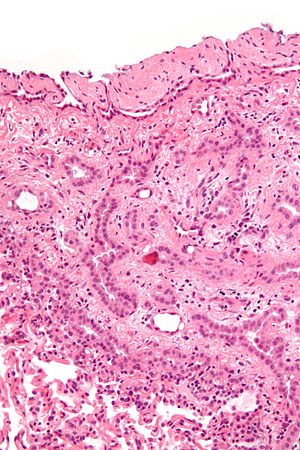
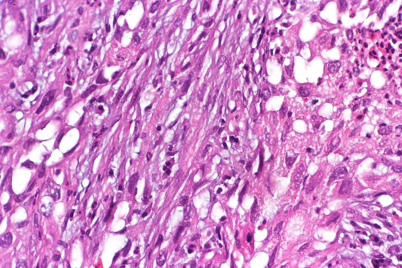
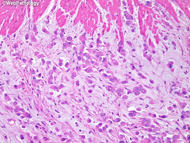
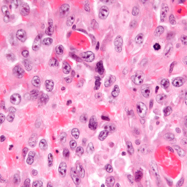
The most common positive stains for pleural mesothelioma are. One highly useful stain is an antibody called pancytokeratin. Malignant mesothelioma mm is an uncommon tumor that can be difficult to diagnose.
Pathology is the study of the causes and effects of disease. Visual survey of surgical pathology with 10802 high quality images of benign and malignant neoplasms related entities. All biopsy and fluid samples are sent to a pathologist.
To provide updated practical guidelines for the pathologic diagnosis of mm. Pathologists examine cells from biopsies to identify cancerous cells types of cancer and the stage. Pathologists involved in the international mesothelioma interest group and others with an interest in the field contributed to this update.
To provide updated practical guidelines for the pathologic diagnosis of mm. Malignant mesothelioma focused malignant mesothelioma with stained slides of pathology. Part of mesothelioma pathology is immunohistochemical staining which helps pathologists determine if mesothelioma is present.
Staining reveals antibodies or proteins also called immunohistochemical markers. The pathology of tumor growth. Calretinin indicated in close to all cases of epithelioid mesothelioma cytokeratin 5 or 56 shown in between 75 and 100 of diagnoses wilms tumor i antigen wt1 expressed in between 70 and 95 of cases.
Malignant mesothelioma mm is an uncommon tumor that can be difficult to diagnose. In the case of mesothelioma immunohistochemistry the most commonly used stain is pan cytokeratin which is often referred to as keratin.
More From Cool Coloring Sheets Hard
- Ema Mesothelioma Pathology Outlines
- Lawyer Agreement Lawyer Inheritance Document Usa
- Princess Dora The Explorer Coloring Pages
- Mesothelioma Commercial Mp3
- Juneau Mesothelioma Law Firm
Incoming Search Terms:
- Histopathology Images Of Mesothelioma Well Differentiated Papillary By Pathpedia Com Pathology E Atlas Mesothelioma Cancer Fighting Foods Food Juneau Mesothelioma Law Firm,
- Pathologic Diagnosis And Classification Of Mesothelioma Springerlink Juneau Mesothelioma Law Firm,
- Nuclear Grade And Necrosis Predict Prognosis In Malignant Epithelioid Pleural Mesothelioma A Multi Institutional Study Modern Pathology Juneau Mesothelioma Law Firm,
- Mesothelioma Pathology Dermnet Nz Juneau Mesothelioma Law Firm,
- Loss Of Expression Of Bap1 Is A Useful Adjunct Which Strongly Supports The Diagnosis Of Mesothelioma In Effusion Cytology Modern Pathology Juneau Mesothelioma Law Firm,
- 2 Juneau Mesothelioma Law Firm,


-Hematoxylin%20&%20Eosin%20Stain-NBP2-77445-img0001.jpg)


