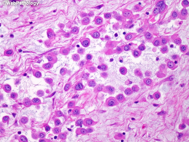
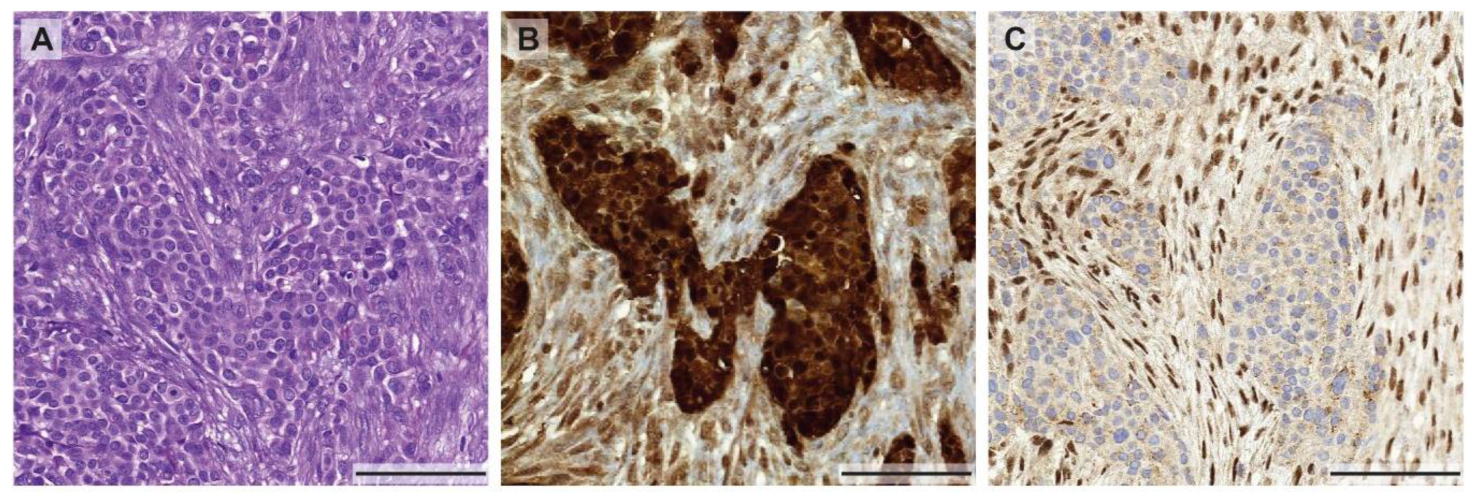
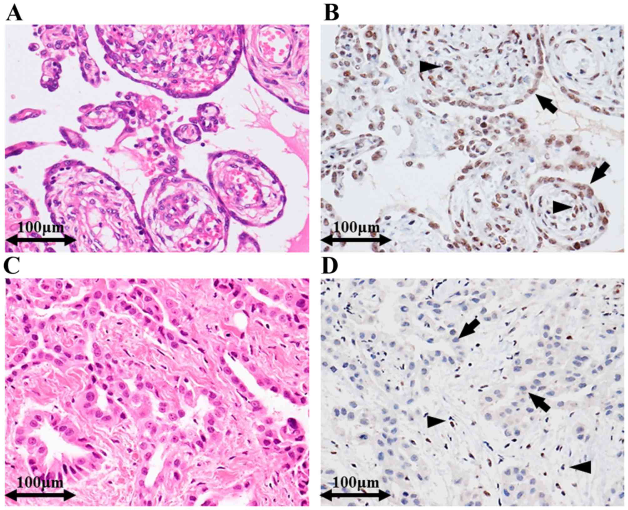
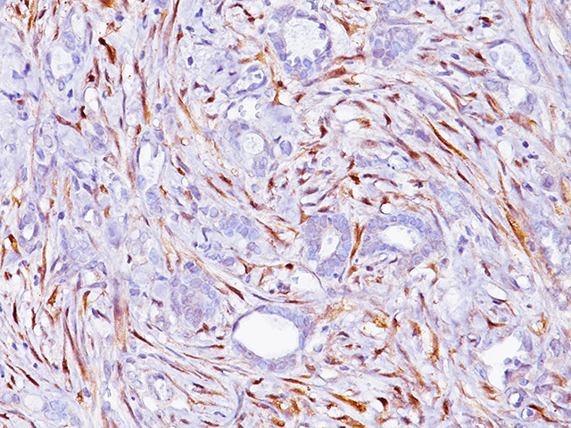
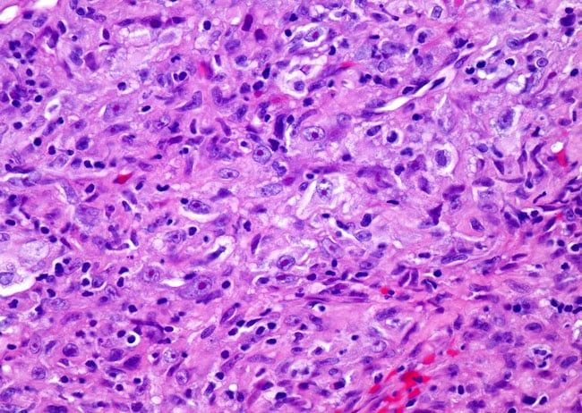
Mesothelioma Staining, Immunohistochemical Staining Of Ctgf Both Mesothelioma Cells And Cafs Download Scientific Diagram
Mesothelioma staining Indeed recently has been hunted by users around us, maybe one of you. People are now accustomed to using the net in gadgets to see video and image data for inspiration, and according to the title of the article I will discuss about Mesothelioma Staining.
- Molecular Drivers Of Mesothelioma
- Utility Of Survivin Bap1 And Ki 67 Immunohistochemistry In Distinguishing Epithelioid Mesothelioma From Reactive Mesothelial Hyperplasia
- Early Detection Of Malignant Pleural Mesothelioma
- Immunohistochemical Staining Of The Mesothelioma Neopl Open I
- Https Patologi Com Guideline 20mesotheliom Pdf
- Mesothelioma Definition Symptoms Diagnosis Treatment Britannica
Find, Read, And Discover Mesothelioma Staining, Such Us:
- Examples For Immunohistochemical Staining Of Malignant Pleural Download Scientific Diagram
- Adam10 Mediates Malignant Pleural Mesothelioma Invasiveness Oncogene
- Representative Ihc Staining For Calretinin A Mesothelioma Cd117 Download Scientific Diagram
- Utility Of Survivin Bap1 And Ki 67 Immunohistochemistry In Distinguishing Epithelioid Mesothelioma From Reactive Mesothelial Hyperplasia
- First Case Report Of Malignant Peritoneal Mesothelioma And Oral Verrucous Carcinoma In A Patient With A Germline Pten Mutation A Combination Of Extremely Rare Diseases With Probable Further Implications Bmc Medical
- Snow Tex 16 Oz
- Plumbers Mesothelioma
- Mark Lanier Attorney Johnson And Johnson
- Fox 5 News Ny
- Leopard Coloring Pages
If you are searching for Leopard Coloring Pages you've arrived at the right place. We have 104 images about leopard coloring pages including images, photos, pictures, wallpapers, and much more. In such web page, we additionally have number of graphics out there. Such as png, jpg, animated gifs, pic art, symbol, blackandwhite, transparent, etc.
8 13 19 in mesothelioma cells ema staining is mainly seen on the cell surfaces but in carcinoma cells ema stains the.
Leopard coloring pages. Bap1 stain also showed utility in the differential of mesothelioma from most common pleural and peritoneal mimickers such as lung and ovary carcinomas with specificity and sensitivity of 9970 and 10070 respectively. This resistance may result in false negatives and delay diagnosis. Positive immunoreactivity for claudin 4 is defined as strong membranous expression with only granular cytoplasmic or very focal staining reported in mesothelioma 79.
Ema is a high molecular weight transmembranous glycosylated protein of the breast mucin complex which is useful for epithelial differentiation and has been found to be present on both carcinoma and mesothelioma cells. The only tumors that have been found to be resistant to the stain are sarcomatoid mesothelioma. Staining can also help pathologists determine which cell type comprises the patients disease.
An antibody is a molecule that binds to another type of molecule. Ema staining in mesothelioma located at the cell. Claudin 4 is now viewed by many expert mesothelioma pathologists and supported by the literature as a superior marker of epithelial differentiation 6 8.
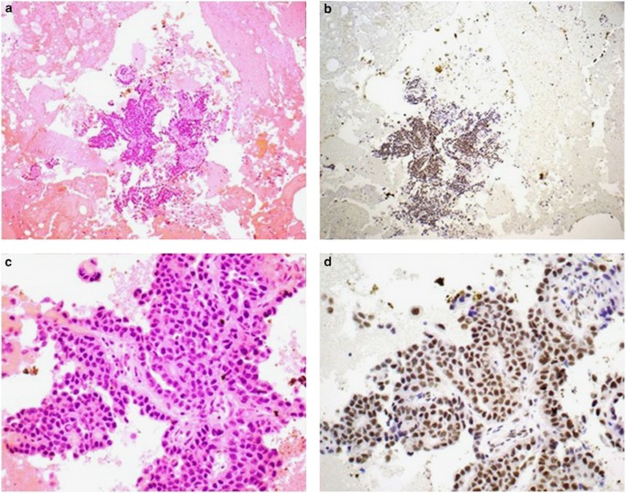
Direct invasion from the lung indirect. The best discriminators among the antibodies considered to be negative markers for mesothelioma are cea moc 31 ber ep4 bg 8 and b723. All mesothelioma biopsycytology pairs showed the same pattern of bap1 or p16 retention or loss in the biopsy and cytology specimens am j surg pathol 201640120126 figure 1.
Pathologists use antibodies designed to stain certain proteins in a color that is easy to see. If a pathologist suspects mesothelioma they will use antibodies to look for proteins that usually occur in mesothelioma cells. Some mesothelioma cell types respond to specific antibodies used in staining.
A panel of four markers two positive and two negative selected based upon availability and which ones yield good staining results in a given laboratory is recommended. The most common positive stains for pleural mesothelioma are.
More From Leopard Coloring Pages
- Mesothelioma Sokolove Law Firm
- Will Lawyers Near Me
- Legal Aid Divorce Attorney
- Mesothelioma Lawyer Mesothelioma Law Firm
- Williams Baker Lawyers
Incoming Search Terms:
- A Cytokeratin And Calretinin Negative Staining Pages 1 6 Text Version Anyflip Williams Baker Lawyers,
- Ijms Free Full Text Heterogeneity In Malignant Pleural Mesothelioma Html Williams Baker Lawyers,
- Malignant Pleural Mesothelioma History Controversy And Future Of A Manmade Epidemic European Respiratory Society Williams Baker Lawyers,
- Pathology Of Mesothelioma Springerlink Williams Baker Lawyers,
- Application Of Immunohistochemistry In Diagnosis And Management Of Malignant Mesothelioma Chapel Translational Lung Cancer Research Williams Baker Lawyers,
- Therapeutic Antitumor Efficacy Of Anti Epidermal Growth Factor Receptor Antibody Cetuximab Against Malignant Pleural Mesothelioma Williams Baker Lawyers,

