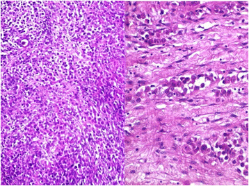
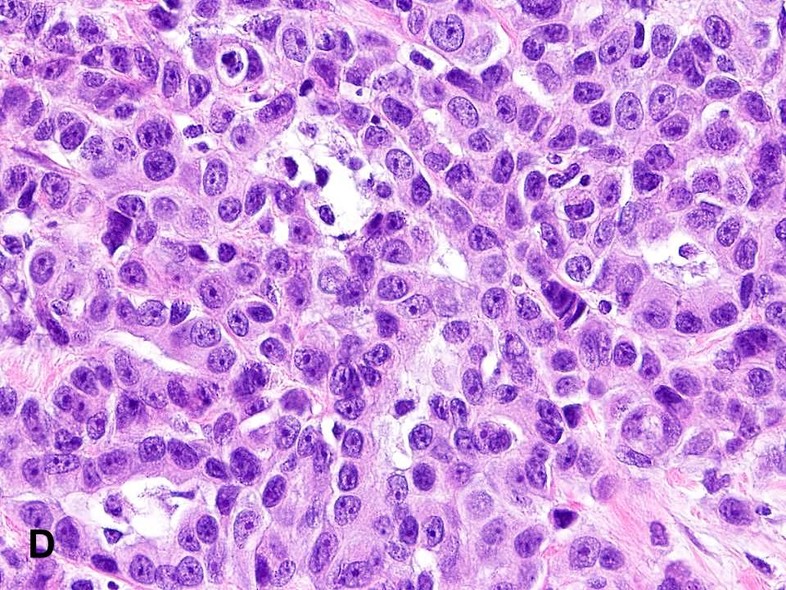
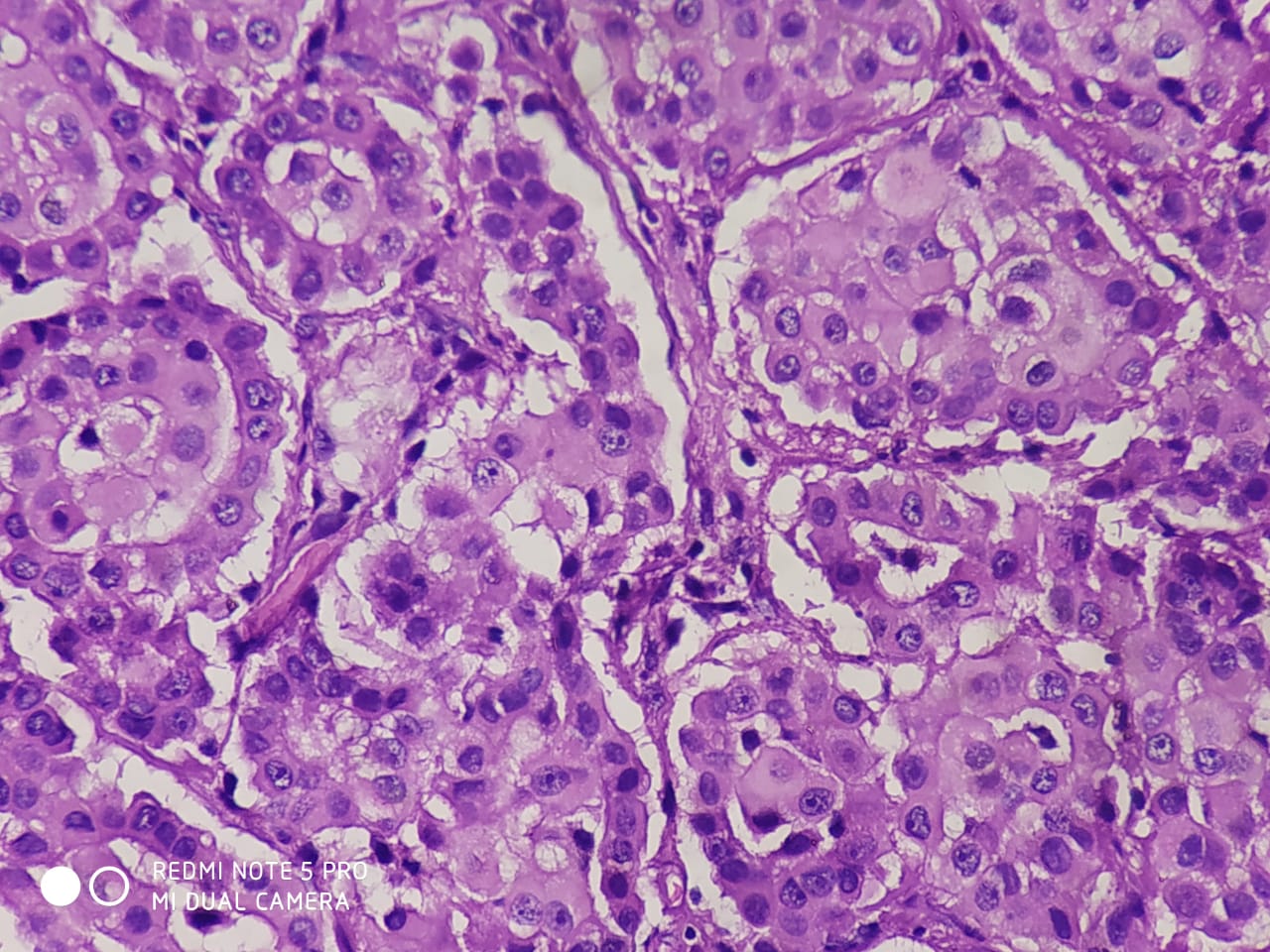
Mesothelioma Malignant Histopathology, Histopathology Result From Fnab Shows Mesothelioma Cell In Mitotic Download Scientific Diagram
Mesothelioma malignant histopathology Indeed recently has been sought by consumers around us, maybe one of you. People are now accustomed to using the net in gadgets to see video and image information for inspiration, and according to the name of the article I will discuss about Mesothelioma Malignant Histopathology.
- Problems In Mesothelioma Diagnosis Addis 2009 Histopathology Wiley Online Library
- Histopathology Of Sarcomatoid Dmm Panels A And B One Of The More Download Scientific Diagram
- Journal Of Pathology And Translational Medicine
- Histopathology Result From Fnab Shows Mesothelioma Cell In Mitotic Download Scientific Diagram
- Malignant Mesothelioma And Other Mesothelial Proliferations Chapter 28 Modern Soft Tissue Pathology
- Pleomorphic Mesothelioma Report Of 10 Cases Modern Pathology
Find, Read, And Discover Mesothelioma Malignant Histopathology, Such Us:
- 2016 Evening Specialty Conference Gynecologic Pathology
- 2016 Evening Specialty Conference Gynecologic Pathology
- Mesothelioma Histology A Study Of Mesothelioma Cells
- P16 Loss And Mitotic Activity Predict Poor Survival In Patients With Peritoneal Malignant Mesothelioma Clinical Cancer Research
- Pleomorphic Mesothelioma Report Of 10 Cases Modern Pathology
- Irvine Divorce Attorney
- Nhs Asbestos
- Coloring Page For Teenager
- Dot A Dot Art Printables
- Malignant Mesothelioma Lawyer
If you re searching for Malignant Mesothelioma Lawyer you've reached the perfect place. We have 104 graphics about malignant mesothelioma lawyer including images, pictures, photos, wallpapers, and much more. In such web page, we additionally have number of graphics out there. Such as png, jpg, animated gifs, pic art, logo, blackandwhite, translucent, etc.

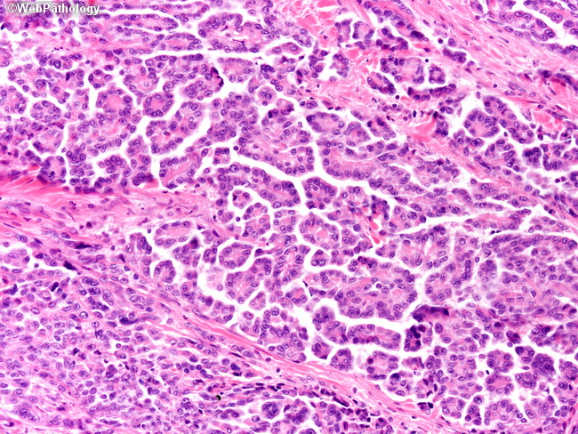
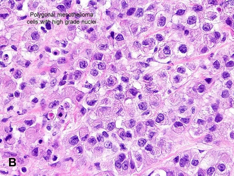
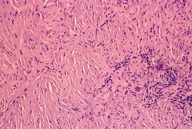
Histopathology Images Of Malignant Mesothelioma By Pathpedia Com Pathology E Atlas Malignant Mesothelioma Lawyer
Confirming that the effusion is malignant.

Malignant mesothelioma lawyer. Malignant mesothelioma is considered biphasic when it contains epithelial and sarcomatoid cells. Each cell type must account for at least 10 of the tumor mass to receive a biphasic diagnosis. Diagnosing malignant mesothelioma through cytology is a two step process.
Mesothelioma has an unusual molecular pathology with loss of tumour supp. The biphasic mixed cell type accounts for 20 to 30 of mesothelioma cases. Mesothelioma histology involves the study of cancerous mesothelial cells.
As a comparable diagnostic tool cytology can be used instead of histopathology to diagnose mesothelioma. In a study of those cases of pleural and peritoneal malignant mesothelioma mm that occurred from 1972 to 1979 occupational histories were obtained during interviews and histopathology of the tumours was reviewed and classified by a member of a mesothelioma. This process is part of mesothelioma pathology which involves examining either tissue or fluid to determine if this cancer exists in the body.
The international mesothelioma interest group has been writing guidelines for pathological diagnosis that are periodically updated. Although the main risk factor is asbestos exposure a virus simian virus 40 sv40 could have a role. Epub 2018 feb 26.
Papillae with myxoid cores each lined by a single mesothelial cell layer invasion is typically not present am j surg pathol 201438990 overall more indolent than peritoneal malignant mesothelioma ann surg oncol 201926852. Malignant mesothelioma in situ. The patient had a history of asbestos exposure and the chest computed tomography scan on initial admission demonstrated an extrapleural sign suggesting a nodular lesion in the chest wall.
Most commonly in the peritoneum rarely pleura and other sites histology. However no nodular lesions were detectable in either of his lungs. We report the autopsy findings of a 58 year old man with malignant mesothelioma in the left pleural cavity.
The guidelines are being updated based on published literature in the last 3 years and experience of more than 20 leading international pathologists in the field who will be. Well differentiated papillary mesothelioma. The los angeles county cancer surveillance program abstracts records on almost all cases of cancer occurring in the county.
Confirming that the cancerous cells began from mesothelial cells. Malignant mesothelioma is an aggressive treatment resistant tumour which is increasing in frequency throughout the world.
More From Malignant Mesothelioma Lawyer
- Melbourne Mesothelioma Attorney
- Christmas Colouring Pdf
- What Is Mesothelioma Lawyer California
- Free Online Coloring Apps For Adults
- Scary Profile Pics
Incoming Search Terms:
- Well Differentiated Papillary Mesothelioma Libre Pathology Scary Profile Pics,
- Webpathology Com A Collection Of Surgical Pathology Images Scary Profile Pics,
- Epithelioid Mesothelioma Histology Scary Profile Pics,
- Mesothelioma Histology A Study Of Mesothelioma Cells Scary Profile Pics,
- Challenges And Controversies In The Diagnosis Of Malignant Mesothelioma Part 2 Malignant Mesothelioma Subtypes Pleural Synovial Sarcoma Molecular And Prognostic Aspects Of Mesothelioma Bap1 Aquaporin 1 And Microrna Journal Of Clinical Pathology Scary Profile Pics,
- Benign And Malignant Mesothelial Proliferation Surgical Pathology Clinics Scary Profile Pics,
