Mesothelioma In A Microscope, Tiny Microscope May Allow Instant Mesothelioma Diagnoses
Mesothelioma in a microscope Indeed recently has been hunted by users around us, maybe one of you personally. Individuals are now accustomed to using the net in gadgets to view video and image data for inspiration, and according to the title of this article I will discuss about Mesothelioma In A Microscope.
- A B Asbestos Bodies Observed Under The Optical Microscope C Download Scientific Diagram
- Pin On Treatment For Mesothelioma
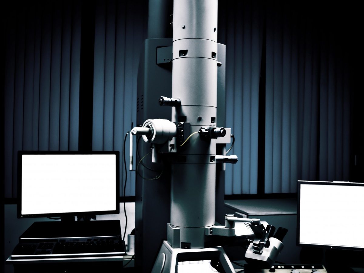
- Diagnosing Mesothelioma With Electron Microscopy Of Cells In Lung Liquid
- Same Cell Correlative Light And Electron Microscopy Of Asbestos Fibers Download Scientific Diagram
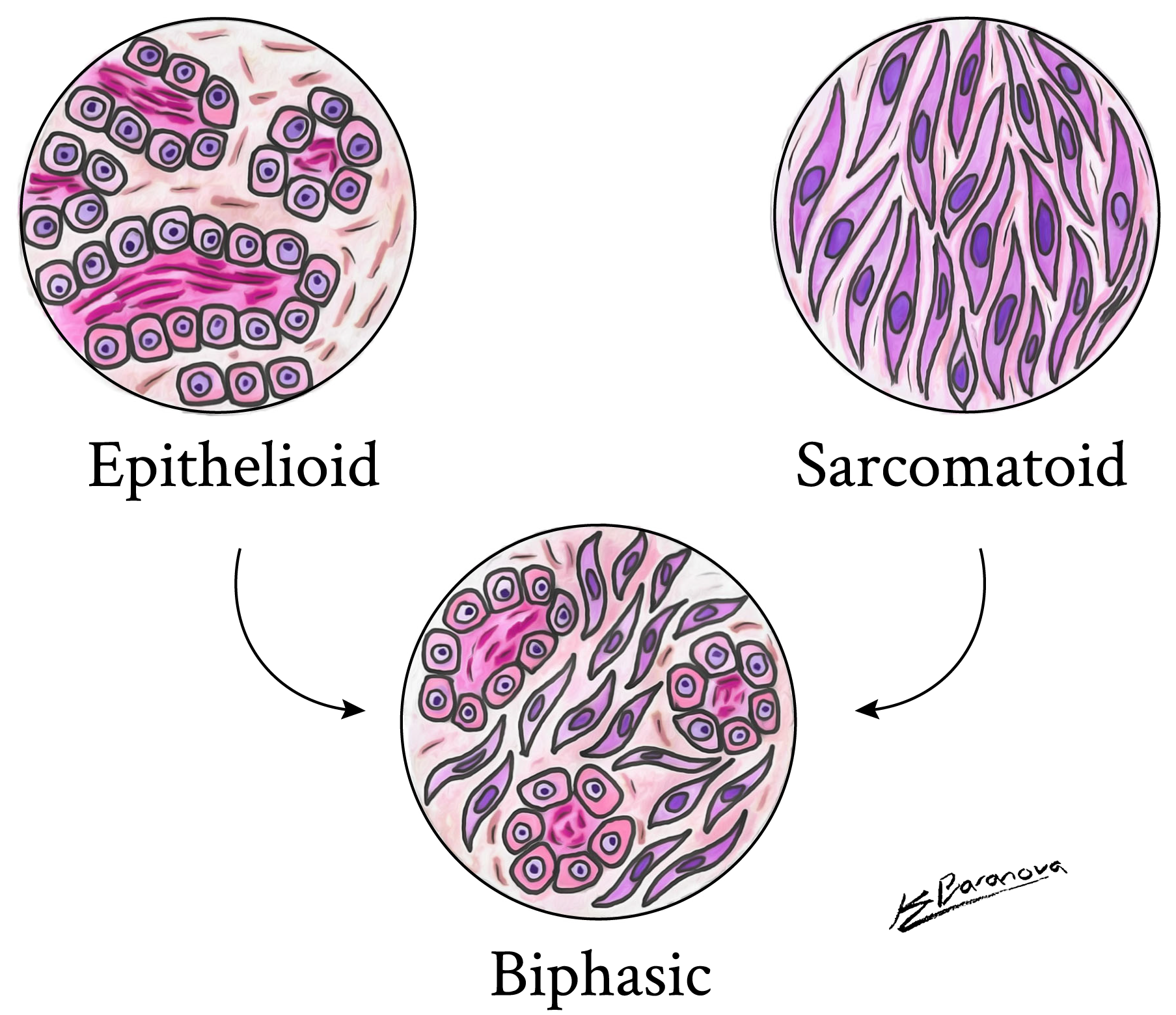
- Epithelioid Mesothelioma The Most Treatable Mesothelioma
- Asbestos Nikon Metrology
Find, Read, And Discover Mesothelioma In A Microscope, Such Us:
- Epithelioid Mesothelioma Treatment Prognosis Diagnosis
- Pin On Sealing Materials Yuanbo
- Coronavirus Postpones Mesothelioma Symposium
- Rcp Launches New Mesothelioma Report Rcp London
- Cytopathology Conference Science Nerd Mesothelioma Cell
- Simmons Citrate Agar Test
- Mermaid Images For Colouring
- Castle Candyland Coloring Pages
- One Third Avenue
- Jack O Lantern Designs Templates
If you re searching for Jack O Lantern Designs Templates you've arrived at the ideal location. We have 100 images about jack o lantern designs templates including pictures, photos, photographs, wallpapers, and more. In such webpage, we additionally provide number of images available. Such as png, jpg, animated gifs, pic art, logo, black and white, transparent, etc.
It is the main procedure used to diagnose pleural mesothelioma and can be taken in two ways.

Jack o lantern designs templates. In the case of mesothelioma immunohistochemistry the most commonly used stain is pan cytokeratin which is often referred to as keratin. Even though a treatment plan for pleural mesothelioma and lung adenocarcinoma may be similar the sooner the diagnosis is made the better the prognosis. Under the microscope mesothelioma may look like other types of cancer.
A biopsy is when a sample of pleural or abdominal tissue is removed for examination under a microscope. For example pleural mesothelioma can resemble some types of lung cancer and peritoneal mesothelioma in women may look like some types of ovarian cancer. This type has smooth thin walled cysts held together by fragile fibrovascular tissue.
Mesothelioma histology or mesothelioma histopathology is the study of tissue for the presence of mesothelioma. This type is also called glandular or microglandular mesothelioma. Via vats video assisted thoracoscopic surgery a type of keyhole surgery.
In this variant of epithelial mesothelioma the cells line small gland like structures. Benign mesothelioma is neither cancerous nor the result of asbestos exposure. The medical term histology refers to the microscopic study of tissue.
Immunohistochemistry works by selecting particular proteins antigens in the tissue sample and seeing what antibodies bind to them under a microscope. Or via ct guided core biopsy which is done under a local anaesthetic using a needle guided by a ct scan.
More From Jack O Lantern Designs Templates
- Corn And Peg Coloring Pages
- Mesothelioma Types Of Asbestos
- Columbia Sc Mesothelioma Lawyer
- Go Fund Me Mesothelioma
- Mesothelioma Study Day
Incoming Search Terms:
- The Cell Structure Of Lung Tissue Affected By The Cancer News Photo Getty Images Mesothelioma Study Day,
- Asbestos Under The Microscope Mesothelioma Study Day,
- Cytopathology Conference Science Nerd Mesothelioma Cell Mesothelioma Study Day,
- Asbestos Detecting Microscope Could Improve Abatement Mesothelioma Com Mesothelioma Study Day,
- Mesothelioma Clinical Trial Testing New Inbrx 109 Antibody Mesothelioma Study Day,
- What To Know About Undergoing A Pleural Mesothelioma Biopsy Mesothelioma Study Day,


