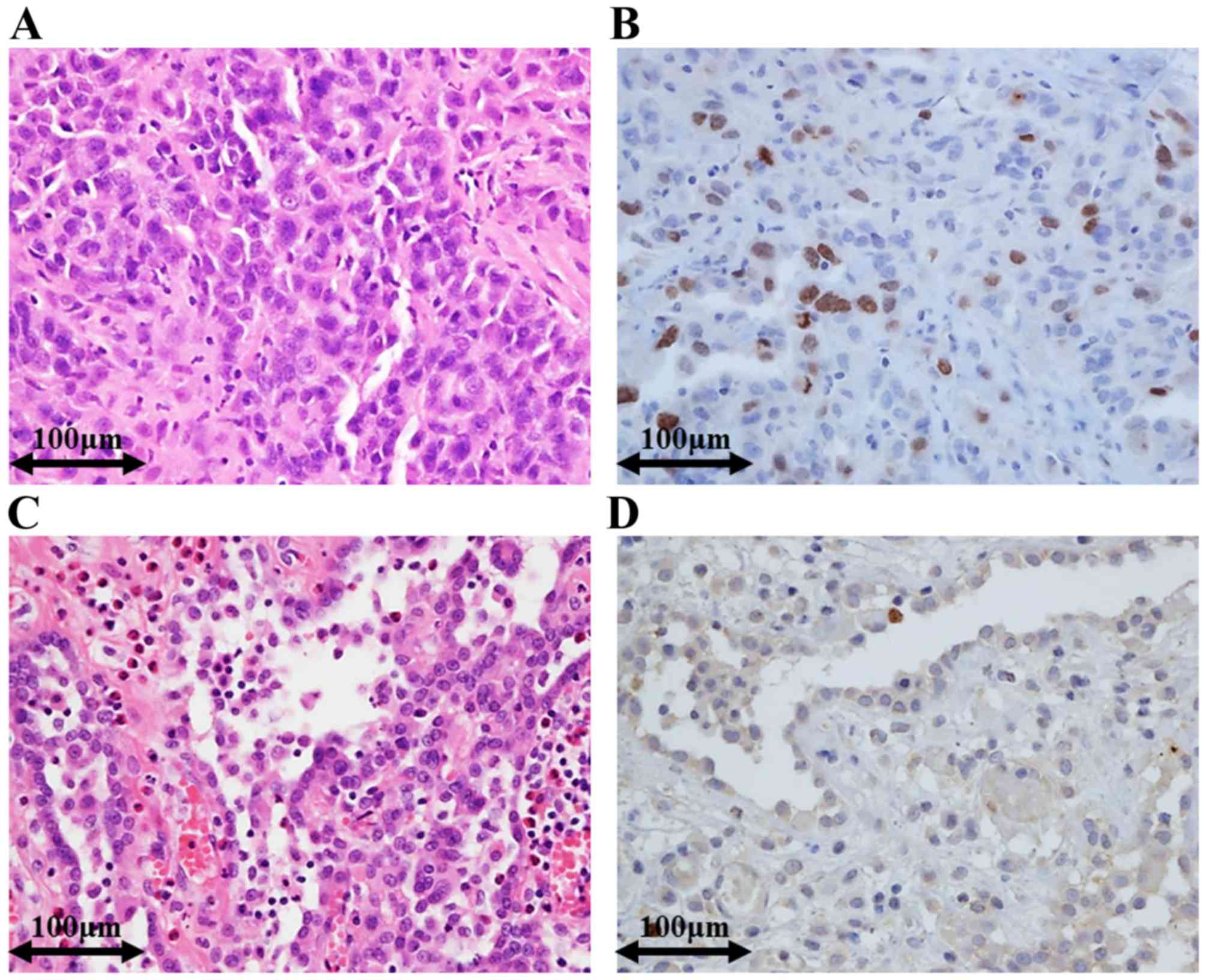
Mesothelioma Ihc Pathology, Pdf The Diagnosis Of Desmopiastic Malignant Mesothelioma And Its Distinction From Fibrous Pleurisy A Histologic And Immunohistochemical Analysis Of 31 Cases Including P53 Immunostaining
Mesothelioma ihc pathology Indeed lately has been sought by users around us, maybe one of you. People now are accustomed to using the internet in gadgets to see image and video data for inspiration, and according to the title of the post I will discuss about Mesothelioma Ihc Pathology.
- Mtap Immunohistochemistry Is An Accurate And Reproducible Surrogate For Cdkn2a Fluorescence In Situ Hybridization In Diagnosis Of Malignant Pleural Mesothelioma Modern Pathology
- Http Phenopath Com Uploads Pdf Mesothelioma Vs Adenocarcinoma Pdf
- Molecular Aspects Of Malignant Mesothelioma And Other Tumors Of The Pleura And Peritoneum Chapter 10 Practical Pathology Of Serous Membranes
- Https Encrypted Tbn0 Gstatic Com Images Q Tbn 3aand9gcrheiekogxvmwle4w4sb4 Bhrsqgcucp5nubatiyl Jo69wqwrt Usqp Cau
- Vasculogenic Mimicry In Malignant Mesothelioma An Experimental And Immunohistochemical Analysis Sciencedirect
- Immunohistochemical Markers Used For Malignant Mesothelioma Diagnosis Download Table
Find, Read, And Discover Mesothelioma Ihc Pathology, Such Us:
- Https Academic Oup Com Ajcp Article Pdf 110 3 397 24884795 Ajcpath110 0397 Pdf
- Https Encrypted Tbn0 Gstatic Com Images Q Tbn 3aand9gcqeiy6h7ynq0bzuozuqljobr7k9gt0qrqzuqzhy7qweho6z2ryw Usqp Cau
- Https Patologi Com Guideline 20mesotheliom Pdf
- 2016 Evening Specialty Conference Pulmonary Pathology
- Https Patologi Com Guideline 20mesotheliom Pdf
- Free Coloring Pictures Of Unicorns
- Captain America Lego Avengers Coloring Pages
- Stage 4 Epithelial Mesothelioma
- Starfish Coloring Pages For Adults
- Multiplication Coloring Worksheets
If you are looking for Multiplication Coloring Worksheets you've arrived at the right location. We have 104 graphics about multiplication coloring worksheets adding pictures, photos, photographs, backgrounds, and more. In such web page, we additionally have variety of graphics out there. Such as png, jpg, animated gifs, pic art, symbol, blackandwhite, translucent, etc.

Bap1 Immunohistochemistry And P16 Fish Results In Combination Provide Higher Confidence In Malignant Pleural Mesothelioma Diagnosis Roc Analysis Of The Two Tests Hida 2016 Pathology International Wiley Online Library Multiplication Coloring Worksheets
Homozygous deletion of 9p21 detected by fluorescence in situ hybridization fish and loss of brca1 associated protein 1 bap1 expression detected by immunohistochemistry ihc are useful for the differentiation between malignant pleural mesothelioma mpm and reactive mesothelial hyperplasia.

Multiplication coloring worksheets. Calretinin is a calcium binding protein that occurs in various types of cells in the body. The role of immunohistochemistry in this differential diagnosis is not as well defined as it is for distinguishing epithelioid mesothelioma from adenocarcinoma. The authors previously described that ihc expression of the protein product of the.
Immunohistochemistry ihc has become an invaluable tool in the differentiation of histological mesothelioma subtypes with the use of antigens which are substances that trigger the production of antibodies by the immune system. 1 department of pathology the university of texas md anderson cancer center houston tx 77030 usa. Differentiating sarcomatoid mesothelioma from other pleural based spindle cell tumours by light microscopy can be challenging especially in a biopsy.
Malignant mesothelioma mm versus other malignant tumors and malignant versus reactive mesothelial proliferations. Overall sensitivity and specificity in mesothelioma is 53 and 98 respectively arch pathol lab med 2012136804 negative stains cd31 positive in angiosarcoma cd34 solitary fibrous tumor s100 neurogenic tumors but mesotheliomas may show focal staining. Of 217 cases circulated among all members of the uscanadian mesothelioma reference panel there was some disagreement about whether the process was benign or malignant in 22 of cases.
Pathology is important to the study of all diseases. This science aids proper diagnosis but also adds to our understanding of how diseases like mesothelioma progress. Contextthe pathologic approach to pleural based lesions is stepwise and uses morphologic assessment correlated with clinical and imaging data supplemented by immunohistochemistry ihc and more recently molecular tests as an aid for 2 main diagnostic problems.
By locating distinct protein antigen markers within mesothelioma tumor cells a diagnosis can be made that is far more accurate than diagnoses using imaging. The distinction between reactive mesothelial hyperplasia mh and malignant mesothelioma mm may be very difficult based only on histologic and morphologic findings. Because of this there is no standard set of markers for mesothelioma.
More From Multiplication Coloring Worksheets
- Mesothelioma Illinois
- Water Cycle Diagram Coloring Page
- Ttt Of Mesothelioma
- Lol Coloring Sheets For Kids
- Disney Pocahontas Coloring Pages
Incoming Search Terms:
- Http Ajcp Oxfordjournals Org Content Ajcpath 112 1 75 Full Pdf Disney Pocahontas Coloring Pages,
- The Pathological And Molecular Diagnosis Of Malignant Pleural Mesothelioma A Literature Review Ali Journal Of Thoracic Disease Disney Pocahontas Coloring Pages,
- Https Www Jto Org Article S1556 0864 19 33232 0 Pdf Disney Pocahontas Coloring Pages,
- Http Ajcp Oxfordjournals Org Content Ajcpath 112 1 75 Full Pdf Disney Pocahontas Coloring Pages,
- Questioning The Prognostic Role Of Bap 1 Immunohistochemistry In Malignant Pleural Mesothelioma A Single Center Experience With Systematic Review And Meta Analysis Lung Cancer Disney Pocahontas Coloring Pages,
- Fatal Hemothorax Caused By Pleural Mesothelioma In A Lion Disney Pocahontas Coloring Pages,
