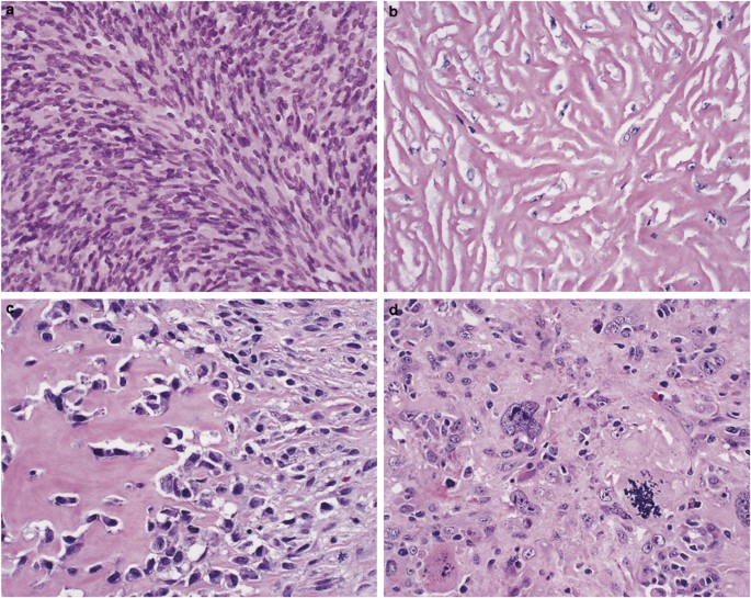
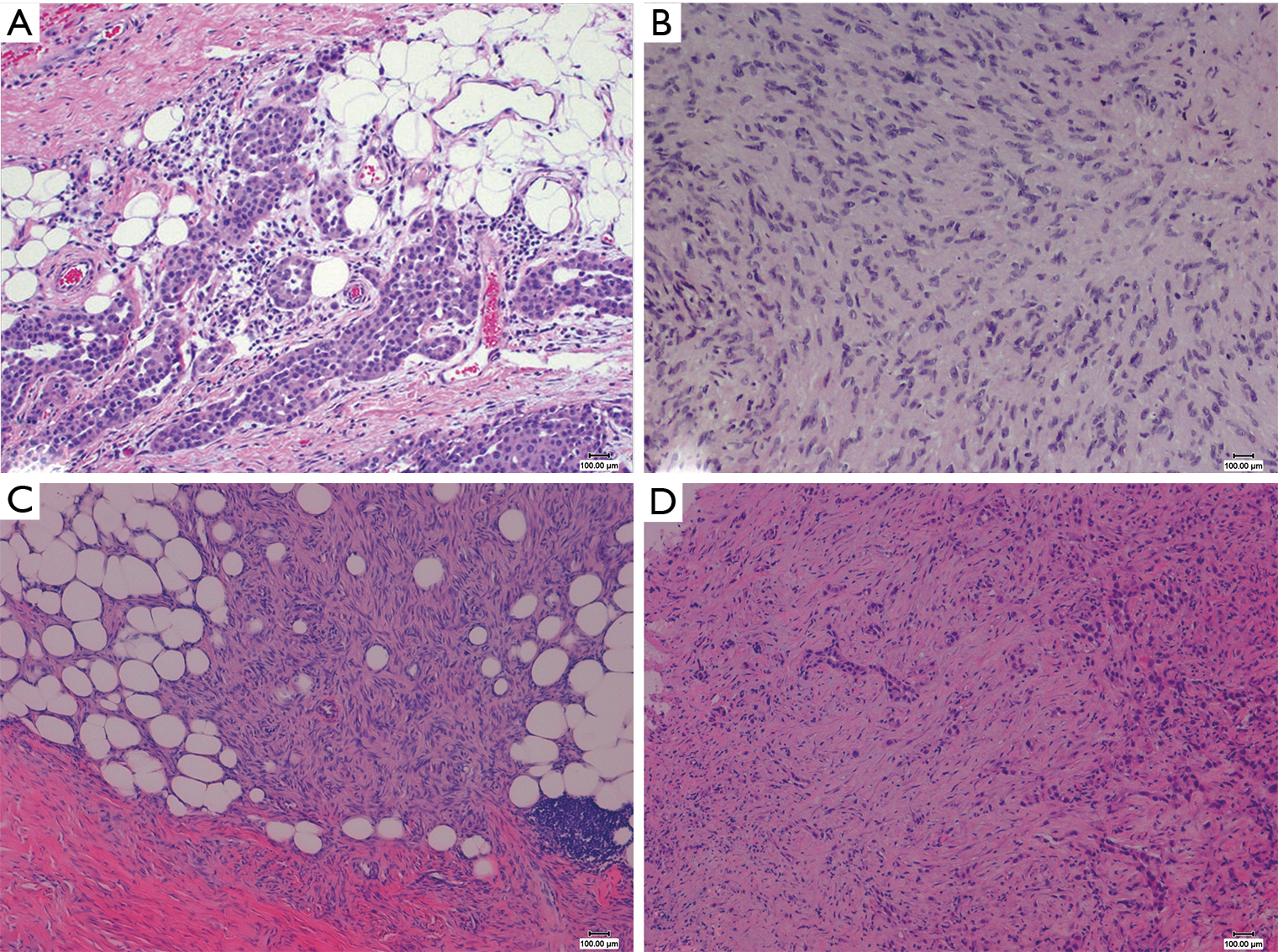
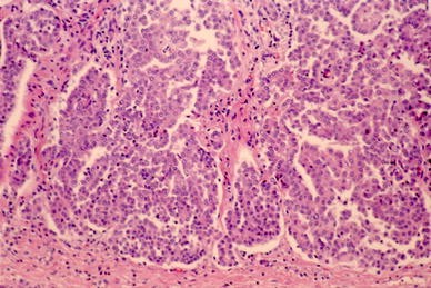
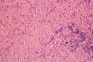
Mesothelioma Histology Images, Shorter Survival In Malignant Pleural Mesothelioma Patients With High Pd L1 Expression Associated With Sarcomatoid Or Biphasic Histology Subtype A Series Of 214 Cases From The Bio Maps Cohort Clinical Lung Cancer
Mesothelioma histology images Indeed lately has been sought by consumers around us, maybe one of you. Individuals now are accustomed to using the net in gadgets to view image and video information for inspiration, and according to the name of this article I will talk about about Mesothelioma Histology Images.
- Malignant Deciduoid Mesothelioma Case Presentation Of An Exceptional Variant And Review Of The Literature Bmc Clinical Pathology Full Text
- Pericardial Mesothelioma Presenting As Chronic Constrictive Pericarditis A Series Of Three Cases From A Single Institution Hui M Harshavardhana K R Uppin Sg Indian J Pathol Microbiol
- Epithelioid Malignant Mesothelioma Thoracic Pathology A Volume In The High Yield Pathology Series Expert Consult Online And Print 1st Edition
- Http Www Ajcr Us Files Ajcr0000001 Pdf
- Histopathology Images Of Malignant Mesothelioma By Pathpedia Com Pathology E Atlas
- Journal Of Pathology And Translational Medicine
Find, Read, And Discover Mesothelioma Histology Images, Such Us:
- Mesothelioma Lungs And Pleura Mypathologyreport Ca Ca
- P16 Loss And Mitotic Activity Predict Poor Survival In Patients With Peritoneal Malignant Mesothelioma Clinical Cancer Research
- Malignant Pleural Mesothelioma Subtypes
- Pathology Of Mesothelioma Springerlink
- Pathology Outlines Mesothelioma Epithelioid
- Iron Spider Coloring Pages Printable
- Printable Coloring Pictures Of Animals
- Epithelial Mesothelioma Survival Rate
- Kids Coloring Pages Paw Patrol
- Dr Nabarro
If you re looking for Dr Nabarro you've reached the ideal location. We have 100 images about dr nabarro adding images, photos, pictures, wallpapers, and much more. In such webpage, we also provide variety of graphics out there. Such as png, jpg, animated gifs, pic art, symbol, blackandwhite, transparent, etc.
Difference between cytology and histology.

Dr nabarro. Malignant epithelial mesothelioma epithelial cells make up the tissues that line the surfaces and cavities of the human body. Papillae with myxoid cores each lined by a single mesothelial cell layer invasion is typically not present am j surg pathol 201438990 overall more indolent than peritoneal malignant mesothelioma ann surg oncol 201926852. There is a well established link between mesothelioma and a sbestos.
With pleural mesothelioma the cancer often spreads to skeletal muscle in the chest cavity. In a malignant epithelial mesothelioma tumor up to 60 of. Some experts have criticized cytology as less effective than histology questioning whether it should be used for mesothelioma diagnosishowever more recent research has determined the two diagnostic procedures to be complementary.
Cytology is less invasive and therefore recommended as the first test for mesothelioma with biopsy recommended if the. Mesothelioma tumors typically present with three distinct histological abnormalities. In peritoneal mesothelioma the tumor often spreads to the liver spleen intestines and other abdominal organs.
It can also spread to deep layers of the skin the diaphragm organs of the abdominal cavity and the lymph nodes. The first test performed by a pathologist is a gross examination which means the tissue sample is reviewed without the use of any tools. Most commonly in the peritoneum rarely pleura and other sites histology.
Well differentiated papillary mesothelioma. Histology samples collected from a biopsy are placed into a container with a preservative and then sent to the pathology lab for examination. Strong association with asbestos exposure either occupational eg shipbuilding construction automotive industry or residential industrial contamination environmental domestic br j ind med 196017260 lung cancer 200445s3 incidence varies by geographic location.
Metastasis to the skin may be the initial presentation and subsequent radiological examinations find the primary tumour in the lung. Mesothelioma is a malignant tumour arises from mesothelial lining of pleura peritoneum pericardium and tunica vaginalis pleural mesothelioma is the most common of these. An important tool used in the definitive diagnosis of disease is histology the microscopic examination of cellular anatomy.
More From Dr Nabarro
- Halloween Things To Do
- Inman Law Firm
- Swear Word Coloring Pages App
- Property Tax Attorney
- Minecraft Skeleton Coloring Pages
Incoming Search Terms:
- Pathology Of Mesothelioma Microscopic Features Dr Sampurna Roy Md Minecraft Skeleton Coloring Pages,
- Epithelioid Mesothelioma Treatment Prognosis Diagnosis Minecraft Skeleton Coloring Pages,
- The Pathological And Molecular Diagnosis Of Malignant Pleural Mesothelioma A Literature Review Ali Journal Of Thoracic Disease Minecraft Skeleton Coloring Pages,
- Webpathology Com A Collection Of Surgical Pathology Images Minecraft Skeleton Coloring Pages,
- Gene Expression Profiling Of Malignant Mesothelioma Clinical Cancer Research Minecraft Skeleton Coloring Pages,
- Early Detection Of Malignant Pleural Mesothelioma Minecraft Skeleton Coloring Pages,


