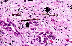
Mesothelioma Histology Description, Pathology Outlines Mesothelioma Epithelioid
Mesothelioma histology description Indeed lately has been hunted by consumers around us, perhaps one of you. Individuals now are accustomed to using the internet in gadgets to view image and video information for inspiration, and according to the name of this article I will talk about about Mesothelioma Histology Description.
- Figure 2 From Reproducibility Of Histological Subtyping Of Malignant Pleural Mesothelioma Semantic Scholar
- Mesothelioma Vs Adenocarcinoma Cytology Creative Art
- Biphasic Mesothelioma Get Treatment And Financial Help
- Pathology Outlines Mesothelioma Epithelioid
- Malignant Peritoneal Mesothelioma In Patients With Endometriosis Journal Of Clinical Pathology
- Webpathology Com A Collection Of Surgical Pathology Images
Find, Read, And Discover Mesothelioma Histology Description, Such Us:
- Pleural Epitheliod Hemangioendothelioma What Started As A Liver Fluke And Ended Up Being Almost Mistaken For Malignant Mesothelioma
- Https Scholarworks Iupui Edu Bitstream 1805 14188 1 Ward 2017 Epithelioid Pdf
- Mesothelioma Oncology Medbullets Step 2 3
- Pleomorphic Mesothelioma Report Of 10 Cases Modern Pathology
- Benign And Malignant Mesothelial Proliferation Surgical Pathology Clinics
- Residual Pleural Thickening
- Nightmare Before Christmas Pumpkin Carving Templates
- Ferrari Colouring Pages Printable
- Halloween Girl
- Meyers Baby Detergent
If you are searching for Meyers Baby Detergent you've reached the ideal place. We have 100 images about meyers baby detergent including images, photos, pictures, wallpapers, and much more. In such webpage, we also have variety of graphics out there. Such as png, jpg, animated gifs, pic art, symbol, black and white, translucent, etc.
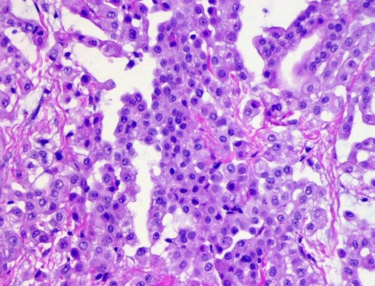
Mesothelioma histology or mesothelioma histopathology is the study of tissue for the presence of mesothelioma.
Meyers baby detergent. Given the presence of the mesothelium in different parts of the body mesothelioma can arise in various locations 17. Pathology reports help doctors diagnose patients and plan treatment. Malignant mesothelioma is a highly aggressive neoplasm of mesothelial cells that usually arises in the pleural cavity but can arise in the peritoneum or pericardium.
Malignant mesothelioma is an invasive and often fatal neoplasm that arises from mesothelium that lines several organs. Common primary sites of origin of mesothelioma are the pleura 8090 and peritoneum 1015 and rarely the pericardium and tunica vaginalis among the three main histologic subtypes of mesothelioma epithelioid tumors are the most common and have a. Mesothelioma is a type of cancer that develops from the thin layer of tissue that covers many of the internal organs known as the mesothelium.
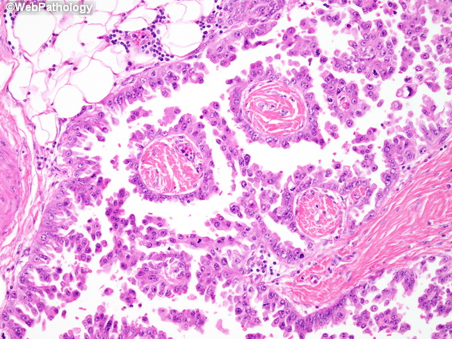
Composed of oval polygonal or cuboidal cells with multiple secondary patterns. This method is used to remove a. Characteristic papillary growth pattern.
The medical term histology refers to the microscopic study of tissue. Doctors typically use one of two methods for a surgical biopsy. Mesothelioma pathology is the study of tissue and fluid samples to determine if infected cells are present.
Mesothelioma pathology is the study of the causes and effects of the disease. The most common area affected is the lining of the lungs and chest wall. This process is part of mesothelioma pathology which involves examining either tissue or fluid to determine if this cancer exists in the body.
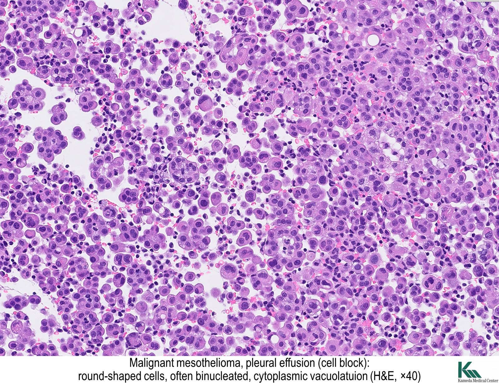
To a mesothelioma pathology report which can be long and complex. A microcystic low power appearance with poor margination and tubules cords of epithelioid cells with characteristic cytoplasmic vacuolation associated with immunosuppressive state in some cases histopathology 2018731013. King md phd in elseviers integrated pathology 2007.
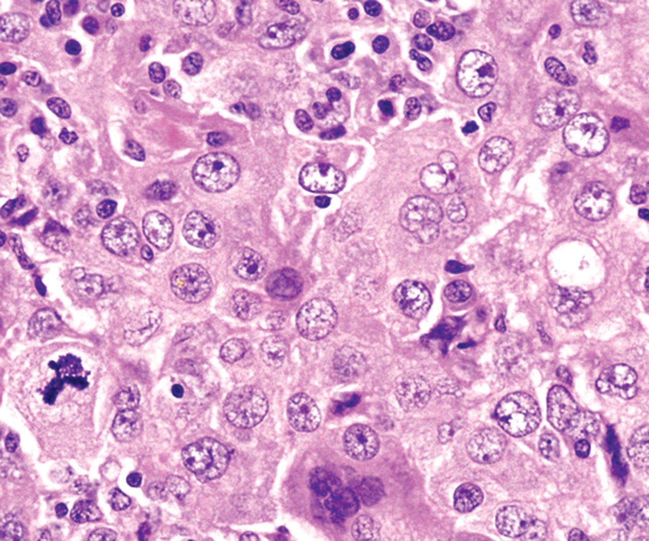
Tumor cells with round nuclei moderate amounts of eosinophilic cytoplasm and conspicuous nucleoli. The most essential parts of the report are the descriptions of the tissue or fluid samples and then the diagnosis section. Elongated tubular structures may also be seen.
Traditional surgical biopsies involve making a large incision in the area where doctors believe cancer exists. Most tumors arise from the pleura and so this article will focus on pleural mesothelioma.
More From Meyers Baby Detergent
- Lawyers For Veterans
- Halloween Pumpkin Pictures
- October Coloring Pages For Adults
- Coloring Pages Christmas Adults
- Free Printable Lol Colouring Pages
Incoming Search Terms:
- Histopathology And Immunoprofile Of The Metastatic Malignant Download Scientific Diagram Free Printable Lol Colouring Pages,
- Journal Of Pathology And Translational Medicine Free Printable Lol Colouring Pages,
- Biotecnol Immunotherapies For Life Free Printable Lol Colouring Pages,
- Webpathology Com A Collection Of Surgical Pathology Images Free Printable Lol Colouring Pages,
- The Pathological And Molecular Diagnosis Of Malignant Pleural Mesothelioma A Literature Review Ali Journal Of Thoracic Disease Free Printable Lol Colouring Pages,
- Application Of Immunohistochemistry In Diagnosis And Management Of Malignant Mesothelioma Chapel Translational Lung Cancer Research Free Printable Lol Colouring Pages,
