Mesothelioma Ct Radiographics, Mesothelioma Radiology Reference Article Radiopaedia Org
Mesothelioma ct radiographics Indeed recently has been sought by consumers around us, perhaps one of you. Individuals now are accustomed to using the internet in gadgets to see image and video data for inspiration, and according to the title of the article I will discuss about Mesothelioma Ct Radiographics.
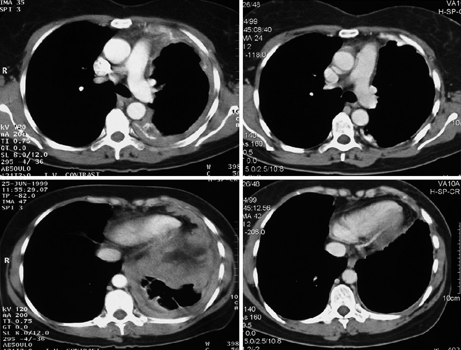
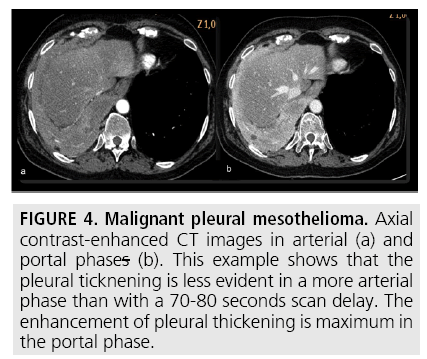
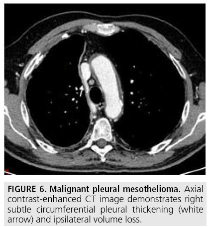
- Malignant Pleural Mesothelioma Evaluation With Ct Mr Imaging And Pet Radiographics
- Malignant Mesothelioma Imaging Overview Radiography Computed Tomography
- Biphasic Malignant Pleural Mesothelioma Masquerading As A Primary Skeletal Tumor
- Mesothelioma Radiology Reference Article Radiopaedia Org
- Imaging Of The Pleura Helm 2010 Journal Of Magnetic Resonance Imaging Wiley Online Library
- Radiological Review Of Pleural Tumors Sureka B Thukral Bb Mittal Mk Mittal A Sinha M Indian J Radiol Imaging
Find, Read, And Discover Mesothelioma Ct Radiographics, Such Us:
- Https Www Resmedjournal Com Article S0954 6111 17 30033 1 Pdf
- Ildulfus39 Mesothelioma Pleural Radiology
- Mesothelioma Radiology Reference Article Radiopaedia Org
- Malignant Mesothelioma Versus Metastatic Carcinoma Of The Pleura A Ct Challenge Iranian Journal Of Radiology Full Text
- Malignant Mesothelioma
- Realistic Grasshopper Coloring Page
- Little Mermaid For Coloring
- Mesothelioma Treatment Cure For Mesothelioma
- Magic Circle Law Firms
- Mesothelioma Ppt
If you re looking for Mesothelioma Ppt you've reached the right place. We ve got 104 graphics about mesothelioma ppt including images, photos, photographs, wallpapers, and more. In such webpage, we additionally have number of graphics out there. Such as png, jpg, animated gifs, pic art, logo, blackandwhite, translucent, etc.

Radiological Review Of Pleural Tumors Sureka B Thukral Bb Mittal Mk Mittal A Sinha M Indian J Radiol Imaging Mesothelioma Ppt
Most tumors arise from the pleura and so this article will focus on pleural mesothelioma.

Mesothelioma ppt. Given the presence of the mesothelium in different parts of the body mesothelioma can arise in various locations 17. Preoperative evaluation of patients with malignant pleural mesothelioma. Mesenteric neoplasms ct appearances of number one and secondary tumors and differential analysis.
Preidler kw steiner h szolar d et al. Malignant pleural mesothelioma mpm is an infrequent neoplasm however mpm is the most common primary malignancy of the pleura and its incidence is estimated as 2000 3000 cases per year in the united states of america 2 3mpm occurs frequently in patients from certain villages in the central and southeastern regions of turkey compared to other parts of the country. Role of integrated ct pet imaging.
Asbestos while the dust settles an imaging evaluate of asbestosrelated ailment. References 1 radiographics 2002. The annual incidence of mpm in the united states is 2500 cases asbestos is a natural silicate material that was used commercially from the late 19th century to the mid 20th century in industries and work environments such as shipyards construction insulation and asbestos.
Cystic appearance of a malignant peritoneal mesothelioma by ultrasonography and computed tomography. Evaluation with ct mr imaging and pet. Computed tomography is the primary imaging modality used for the diagnosis and staging of mpm.
7 wang zj reddy gp gotway mb et al. J thorac imaging 2006. 12a 12b 12c 12d and 13a 13b differentiating well differentiated papillary mesothelioma from tuberculous peritonitis serous surface papillary carcinoma or peritoneal.
Mesothelioma also known as malignant mesothelioma is an aggressive malignant tumor of the mesothelium. However when these tumors present with nonspecific ct findings such as peritoneal thickening multiple peritoneal nodules omental infiltration and ascites figs. Pleural mesothelioma 90 covered in this article.
Mpm most commonly occurs in patients aged 5070 years and is more common in men than in women with a ratio of 41. Doctors use imaging scans such as x rays ct scans mris and others as noninvasive tools that help detect tumors in the body when a patient experiences symptoms usually associated with an asbestos related disease such as mesothelioma. 8 truong mt marom em erasmus jj.
Radiology information training carrier. Imaging plays an essential role in the evaluation of malignant pleural mesothelioma mpm. Eur j radiol 1994.
Mesothelioma is a malignant neoplasm originating from pleural or peritoneal surfaces. Mesenteric neoplasms ct appearances of primary and secondary.
More From Mesothelioma Ppt
- Mesothelioma Spread To Lungs
- Trippy Alien Coloring Pages
- Lawyer Letter For Debt Collection
- Copyright Lawyer Nyc
- Multicystic Peritoneal Mesothelioma A Case
Incoming Search Terms:
- Malignant Pleural Mesothelioma Evaluation With Ct Mr Imaging And Pet Radiographics Multicystic Peritoneal Mesothelioma A Case,
- Https Www Resmedjournal Com Article S0954 6111 17 30033 1 Pdf Multicystic Peritoneal Mesothelioma A Case,
- Overview Of Treatment Related Complications In Malignant Pleural Mesothelioma Murphy Annals Of Translational Medicine Multicystic Peritoneal Mesothelioma A Case,
- Malignant Pleural Mesothelioma Presenting As Perifissural Nodules Radiology Case Radiopaedia Org Multicystic Peritoneal Mesothelioma A Case,
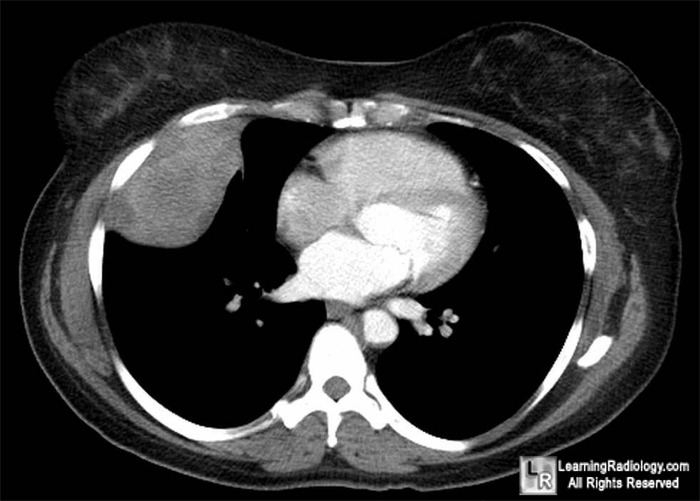
- Learningradiology Localized Fibrous Tumor Pleura Solitary Benign Mesothelioma Pleural Fibroma Radiology Multicystic Peritoneal Mesothelioma A Case,
- Mesothelioma Radiology Case Radiopaedia Org Multicystic Peritoneal Mesothelioma A Case,


