Mesothelioma Abdomen Radiopaedia, Senildi47 Benign Mesothelioma Radiopaedia
Mesothelioma abdomen radiopaedia Indeed lately is being hunted by users around us, maybe one of you personally. People are now accustomed to using the net in gadgets to view video and image information for inspiration, and according to the name of the post I will talk about about Mesothelioma Abdomen Radiopaedia.
- Peritoneal Metastases Radiology Reference Article Radiopaedia Org
- Peritoneal Metastases Radiology Reference Article Radiopaedia Org
- Senildi47 Benign Mesothelioma Radiopaedia
- The Radiology Assistant Peritoneal Pathology
- Mesothelioma Xray
- Pleural Plaques Causes Symptoms Diagnosis Treatment
Find, Read, And Discover Mesothelioma Abdomen Radiopaedia, Such Us:
- Malignant Peritoneal Mesothelioma Radiology Reference Article Radiopaedia Org
- Mesothelioma Radiology Reference Article Radiopaedia Org
- The Grinstaff Group Part 2 Mesothelioma College Of Engineering
- Senildi47 Benign Mesothelioma Radiopaedia
- Sarcoidosis Radiology Case Radiopaedia Org Radiology Radiology Imaging Internal Medicine
- How Much Compensation Is A Mesothelioma Claim
- Crayola Coloring Pages Unicorn
- Hot Air Balloon Coloring Pages
- Fairytale Castle Coloring Pages
- Second Hand Mesothelioma
If you re looking for Second Hand Mesothelioma you've arrived at the right location. We have 104 graphics about second hand mesothelioma adding pictures, pictures, photos, backgrounds, and much more. In these page, we also have variety of graphics out there. Such as png, jpg, animated gifs, pic art, logo, blackandwhite, transparent, etc.
Primary peritoneal tumors are uncommon lesions that arise from the mesothelial or submesothelial layers of the peritoneum.

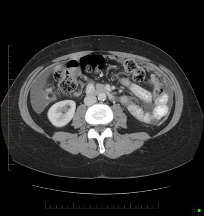
Second hand mesothelioma. Peritoneal mesothelioma in a 73 year old woman with biopsy proved malignant mesothelioma. Peritoneal mesothelioma is very similar to peritoneal carcinomatosis but usually no primary neoplasm is known peritoneal sarcomatosis. Malignant peritoneal mesothelioma is an uncommon primary tumor of the peritoneal lining.
Catch up on exam 1 now and sit exam 2 this. B sagittal us image of the lower abdomen shows two small hypoechoic implants in the near field arrows. Is also known as intraabdominal fibromatosis abdominal desmoid or desmoid tumor.
Sarcoma most commonly metastases from a gastrointestinal sarcoma 1. It shares epidemiological and pathological features with but is less common than its pleural counterpart which is described in detail in the general article on mesothelioma. It is a locally aggressive tumor which often recurs but does not metastasize.
Primary malignant mesothelioma multicystic mesothelioma primary peritoneal serous carcinoma leiomyomatosis peritonealis disseminata and desmoplastic small round cell tumor are the most prominent of these rare lesions. If the primary tumor is of mesenchymal origin ie. It can have a myxoid stroma resulting in a low attenuation on ct and a high attenuation on t2wi.
No pleural plaques were seen on the chest ct scan. Extensive ascites including interloop fluid with enhancing greater omental infiltration. In this case the initial histological diagnosis of metastatic adenocarcinoma was revised following review of clinical and radiologal features and the correct diagnosis was confirmed with special stains calretinin ck56 mesothelin and wt1 which are.
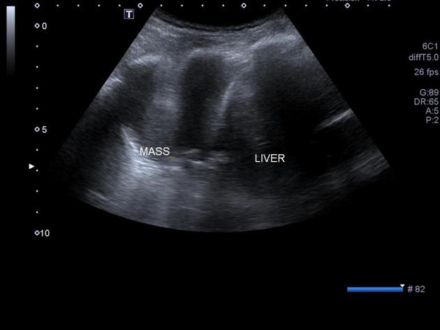
The differentiation between adenocarcinoma and mesothelioma can be difficult at histology. A sagittal us image of the left upper quadrant shows a lobulated heterogeneous mass m that involves the greater omentum. Reference article this is a summary.
This is a basic article for medical students and other non radiologists bowel perforation is an acute surgical emergency where there is a release of gastric or intestinal contents into the peritoneal space. Radiopaedias frcr 2b reporting practice exams. A low signal.
Multiple liver lesions all show nodular peripheral enhancement.
More From Second Hand Mesothelioma
- Free Printable Unicorn Coloring Pages For Kids
- Slip And Fall Attorney New York
- Hardie Board Asbestos
- Ebv Mesothelioma
- What To Do After Being Exposed To Asbestos
Incoming Search Terms:
- X6hw Zfpjk0bmm What To Do After Being Exposed To Asbestos,
- Ct Imaging Of Peritoneal Carcinomatosis And Its Mimics Sciencedirect What To Do After Being Exposed To Asbestos,
- Mesothelioma Radiology Reference Article Radiopaedia Org What To Do After Being Exposed To Asbestos,
- 100 Mejores Imagenes De Patologia De Ct Scan Patologia Radiologia Imagenologia What To Do After Being Exposed To Asbestos,
- Mesothelioma Radiology Reference Article Radiopaedia Org What To Do After Being Exposed To Asbestos,
- Malignant Peritoneal Mesothelioma In A Patient With Intestinal Fistula Incisional Hernia And Abdominal Infection A Case Report What To Do After Being Exposed To Asbestos,


