Mesothelial Cells Pathology Outlines, Neuroendocrine Cancer Pathology Outlines Recent Posts
Mesothelial cells pathology outlines Indeed recently is being hunted by consumers around us, maybe one of you personally. People now are accustomed to using the internet in gadgets to view video and image data for inspiration, and according to the name of this article I will discuss about Mesothelial Cells Pathology Outlines.
- Pathology Outlines Mesothelioma Epithelioid
- Neuroendocrine Cancer Pathology Outlines Recent Posts
- Pathology Outlines Hernia Sac With Mesothelial Entrapment
- Pathology Outlines Mesothelioma Epithelioid
- Adenom Parotid Icd 9 Neuroendocrine Cancer Pathology Outlines
- Pathology Outlines Diffuse Malignant Mesothelioma
Find, Read, And Discover Mesothelial Cells Pathology Outlines, Such Us:
- Pathology Outlines Mesothelial
- Reactive Mesothelial Cells Can Exhibit Coarse Chromatin Irregular Nuclear Outlines And Nucleoli It Is Important To Microscopic Cells Medical Laboratory Cell
- Squamous Cell Papilloma Skin Pathology Outlines Virus Bucuresti
- Pdf Peritoneal Washing Cytology
- Neuroendocrine Cancer Pathology Outlines Recent Posts
- New York Lawyer Aesthetic
- Fourth Of July Coloring Pages
- Weed Costume
- Mesothelioma Treatment Small Incision
- Law Firms In Bellville
If you re searching for Law Firms In Bellville you've arrived at the ideal place. We have 104 graphics about law firms in bellville including images, pictures, photos, wallpapers, and more. In these webpage, we also provide number of images out there. Such as png, jpg, animated gifs, pic art, symbol, blackandwhite, translucent, etc.
This review discusses some of the functions of mesothelial cells regarding maintenance of serosal integrity and outlines the mechanisms involved in mesothelial healing.

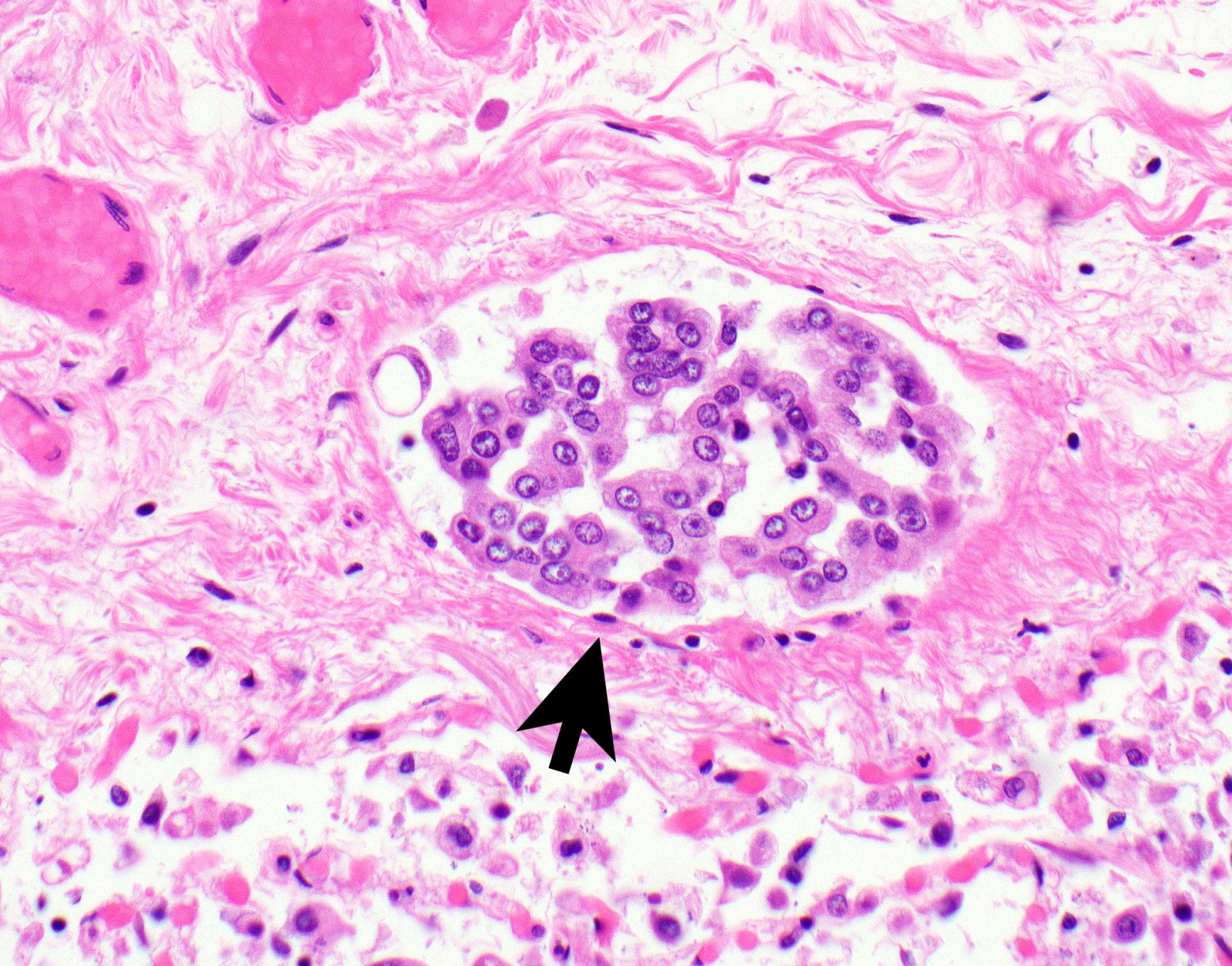
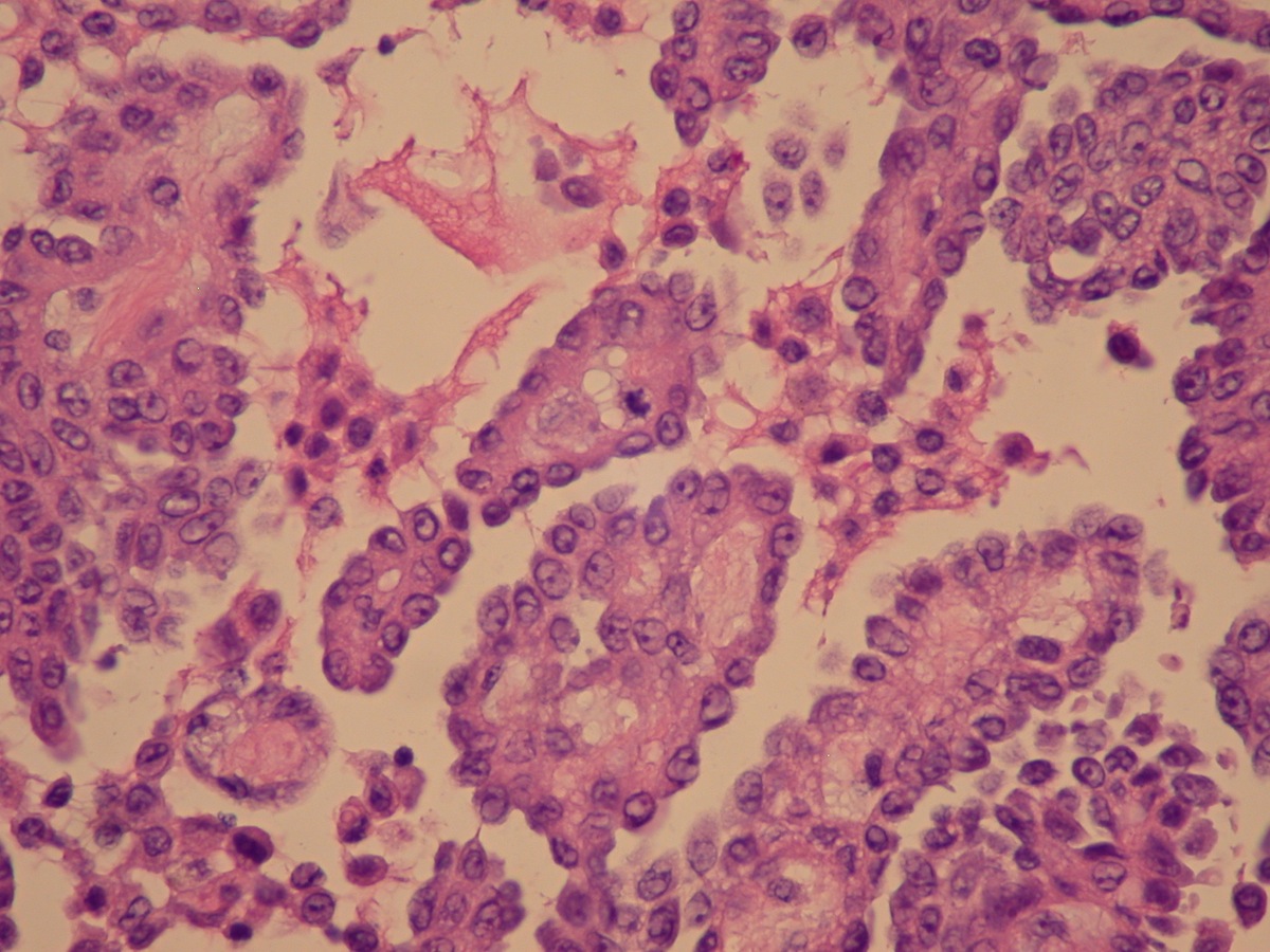
Law firms in bellville. Pathology revealed a cystic mass like lesion measuring 3 3cm in diameter and hernia sac. Usually mesothelial cells will be numerous dispersed or present in small clusters clusters of 12 cells is unusual in simple hyperplasia binucleation multinucleation mitosis prominent nucleolus can be seen in benign proliferations two or more mesothelial cells are often separated by window or a narrow space. It may also include repopulation from free floating mesothelial cells or possibly.
The healing of disrupted serosa includes the multiplication and migration of mesothelial cells from the edges of the injured area. The hernia sac was adherent to the medial aspect of the cyst. Usually mesothelial cells will be numerous dispersed or present in small clusters clusters of 12 cells is unusual in simple hyperplasia binucleation multinucleation mitosis prominent nucleolus can be seen in benign proliferations two or more mesothelial cells are often separated by window or a narrow space.
Mesothelioma pathology provides a full picture of the cancer contributing to a more accurate diagnosis and an informed treatment plan. Adipocytes hum pathol 200637312 endometrial stromal cells leydig cells of testis mast cells mesothelial cells nerves good positive control nordic ovarian theca lutein and theca interna cells sertoli cells hum pathol 200334994. In addition the pathogenesis of adhesion formation is discussed with particular attention to the potential role of mesothelial cells in both preventing and inducing their.
When irritated or injured the mesothelial lining of the peritoneal cavity can show focal or diffuse hyperplasiasome of the situations where mesothelial hyperplasia may be encountered include. Histopathologic examination revealed simple mesothelial cyst with chronic inflammation lined by regular mesothelial cells without no atypia or mitosis. Mesothelial hyperplasia represents a normal reaction to injury.
The article deals with cytopathology specimens from spaces lined with mesothelium ie. Pathologists examine cells from biopsies to identify cancerous cells types of cancer and the stage. The pathology of tumor growth.
Mesothelial cytopathology is a large part of cytopathology. Weiss in modern surgical pathology second edition 2009. Hernia sacs especially in children hydroceles cirrhosis collagen vascular diseases infections long standing effusion of any cause acute appendicitis and ruptured ectopic pregnancy.
More From Law Firms In Bellville
- Hatchimals Coloring Pages Unicorn
- Free Coloring Pages For Teens
- Shapes Coloring
- Chattanooga Mesothelioma Claim
- Cute Hedgehog Coloring Pages
Incoming Search Terms:
- Urethral Papilloma Pathology Outlines Curs Engleza Partea 2 Corectat Lari Cute Hedgehog Coloring Pages,
- Pathology Outlines Diffuse Malignant Mesothelioma Cute Hedgehog Coloring Pages,
- Pathology Outlines Pas Periodic Acid Schiff Cute Hedgehog Coloring Pages,
- Pdf Peritoneal Washing Cytology Cute Hedgehog Coloring Pages,
- Pathology Outlines Mesothelial Cute Hedgehog Coloring Pages,
- Pathology Outlines Diffuse Malignant Mesothelioma Cute Hedgehog Coloring Pages,


