Irregular Pleural Thickening, Benign Pleural Thickening Radiology Key
Irregular pleural thickening Indeed lately is being sought by users around us, maybe one of you. People now are accustomed to using the internet in gadgets to view video and image data for inspiration, and according to the name of this article I will discuss about Irregular Pleural Thickening.
- Thorax 8 2 Pleural Space Case 8 2 2 Benign And Malignant Pleural Thickening Ultrasound Cases
- Pleural Thickening Plaque Radio Pandora Screenshot
- Malignant Pleural Effusion Pulmonology Advisor
- Features Of Lung Ultrasound In Covid 19 Infection Critical Care Sonography
- Presentation1 Ultrasound Examination Of The Chest
- Radiological Imaging Of Pleural Diseases
Find, Read, And Discover Irregular Pleural Thickening, Such Us:
- Pleural Plaques Causes Symptoms Diagnosis Treatment
- Epos Trade
- Http Pdf Posterng Netkey At Download Index Php Module Get Pdf By Id Poster Id 105229
- Pleural Thickening Radiology Reference Article Radiopaedia Org
- Https Jksronline Org Synapse Data Pdfdata 1016jkrs Jkrs 26 735 Pdf
- Queens Car Accident Attorney
- Twisty Noodle Letter E
- Georgia Bar Member Search
- If You Or A Loved One Mesothelioma
- When Was Asbestos Found To Be Dangerous
If you are searching for When Was Asbestos Found To Be Dangerous you've come to the ideal place. We ve got 104 graphics about when was asbestos found to be dangerous adding images, photos, pictures, wallpapers, and much more. In these webpage, we also have variety of graphics out there. Such as png, jpg, animated gifs, pic art, symbol, blackandwhite, transparent, etc.
Https Encrypted Tbn0 Gstatic Com Images Q Tbn 3aand9gcra1 Jjcnemaqcogkly56taklchvofjonh6m9rnxpel8y3koja7 Usqp Cau When Was Asbestos Found To Be Dangerous

Pleural Plaques Diffuse Pleural Thickening Rounded Atelectasis And Download Scientific Diagram When Was Asbestos Found To Be Dangerous
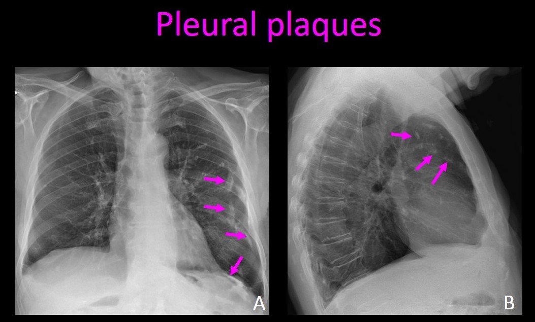
Adjacent to costal pleural surfaces located 1 cm from the pleura according to some publications 4.

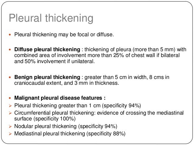
When was asbestos found to be dangerous. Pleural thickening happens when the tissue layers that cover the lungs known as pleura become thickened. Infections from bacterial pneumonia. Benign pleural thickening caused by fibrosis is the second most common pleural abnormality the most common one being effusion.
It can occur with both benign and malignant pleural disease. Diffuse pleural thickening dpt. Benign or malignant tumor.
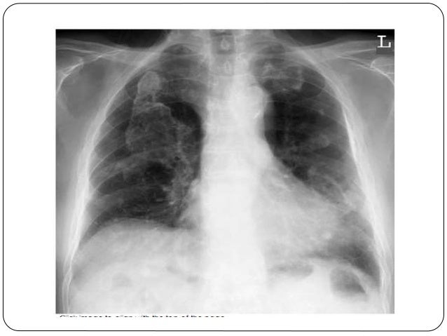
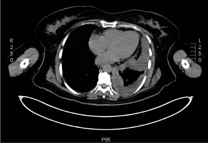
Pleural thickening can be seen on computed tomography scans. Pericardial pleural thickening looked ir regular is called by a shaggy cardiac silhouette and the surface of the diaphragm which is looked irregular and non slippery edges is called by ill de fined diaphragmatic contours. Subpleural reticulation is a type of reticular interstitial pattern where the changes are typically in a peripheral subpleural distribution ie.
Pleural thickening is a common side effect of exposure to asbestos and this thickening is an early warning sign for diagnosis. Non malignant pleural diseases affecting the outer lining of the pleura diffuse pleural thickening dpt and pleural plaques. Thickening of 50 or more of either the left or right pleura.
According to etiology it may be classified as. Pleural fibrosis has a number of causes and is the outcome of many pleural diseases and a potential complication of every inflammatory condition that affects the lungs. As a result the lungs do not expand freely and breathing will then be labored.
Thickening confined to one or more specific areas of the pleura. It can arise in a number of pathological situations as well as in certain physiological situations. The condition is usually first spotted through a chest x ray in which pleural thickening appears as an irregular shadow of the pleura.
The pleura show a variety of patterns of fibrosis. Pleural thickening may be divided into two basic groups. The most common cause of this pathology is asbestosis.
Causes of pleural thickening include. This type is benign unless the pleura has thickened more than two centimeters. Ct scans can be used to diagnose pleural plaques and asbestosis.
Circumferential and pleural wall thickening is more than 1 cm. The pleural membranes thicken as a result of the chronic irritation and inflammation that asbestos fibers cause when they lodge in the lungs. Asbestos fibers are so small they cannot be seen with the naked eye but in.
Thickening of the top most portion of the pleura. Dpt may also be diagnosed in a patient with. Ct scans can detect early signs of pleural thickening when scar tissue is.
More From When Was Asbestos Found To Be Dangerous
- Santa Claus Coloring Pages Online
- How To Prevent Malignant Mesothelioma
- Dora Boots Coloring Pages
- Coloring Book Info Coloring Pages
- Therapeutic Coloring Pages
Incoming Search Terms:
- Benign Pleural Thickening Radiology Key Therapeutic Coloring Pages,
- Https Encrypted Tbn0 Gstatic Com Images Q Tbn 3aand9gct5wagysvxv6irl7dgfam2 Tlmenkhg96a8naqacd 7ymwnujus Usqp Cau Therapeutic Coloring Pages,
- Benign Pleural Thickening Radiology Key Therapeutic Coloring Pages,
- Pleural Thickening On Screening Chest X Rays A Single Institutional Study Respiratory Research Full Text Therapeutic Coloring Pages,
- Clinmed International Library The Many Faces Of Lung Cancer International Journal Of Cancer And Clinical Research Therapeutic Coloring Pages,
- Pin On Pleural Ultrasound Therapeutic Coloring Pages,


