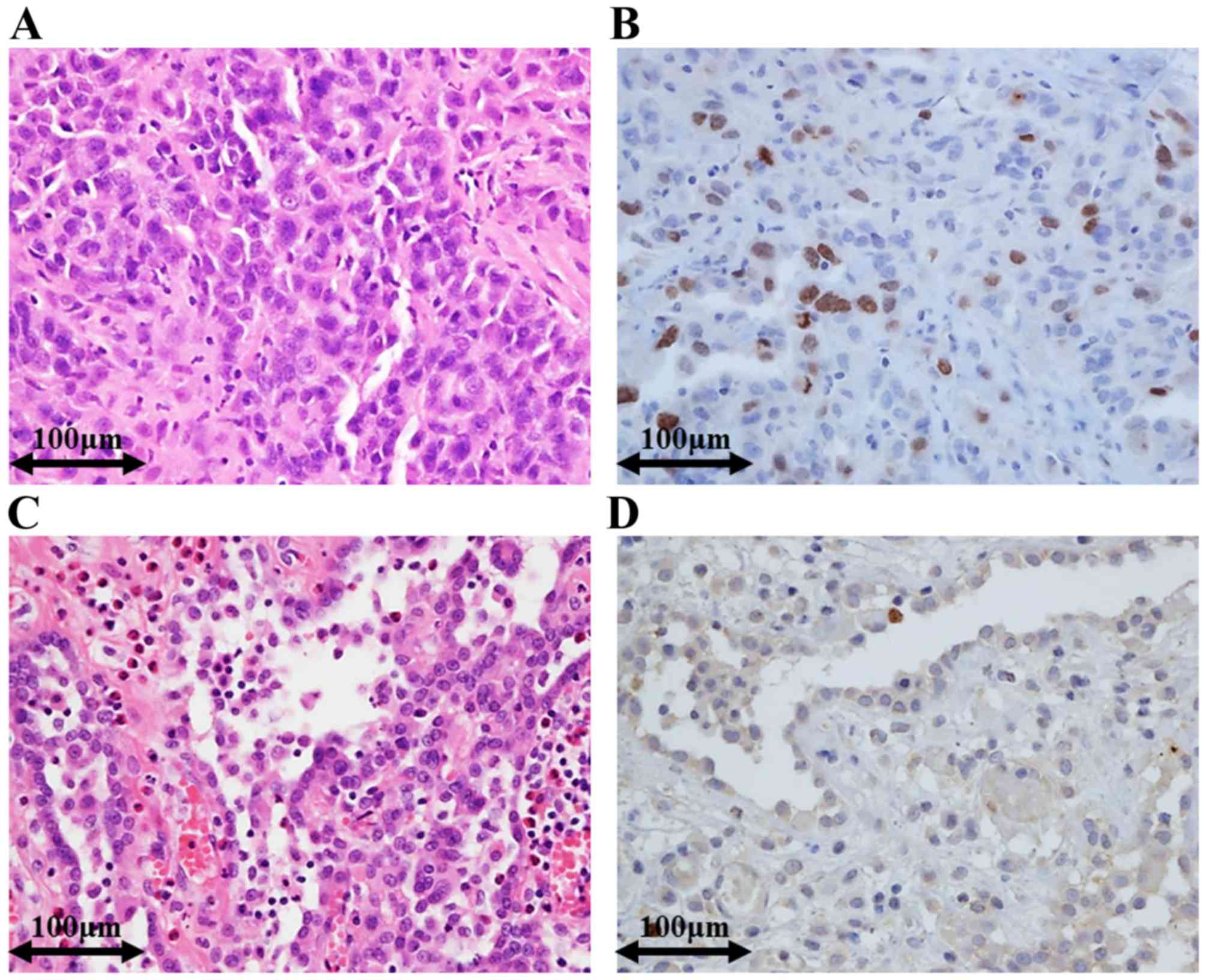
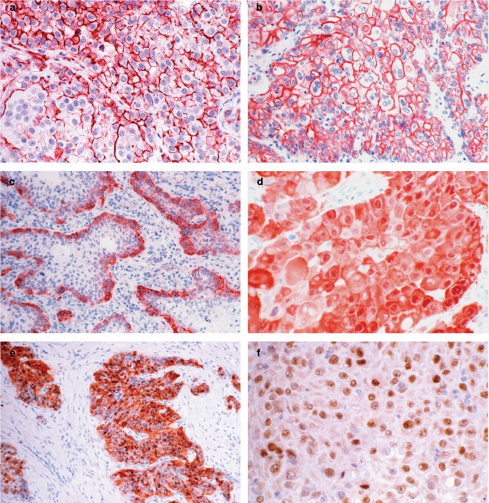
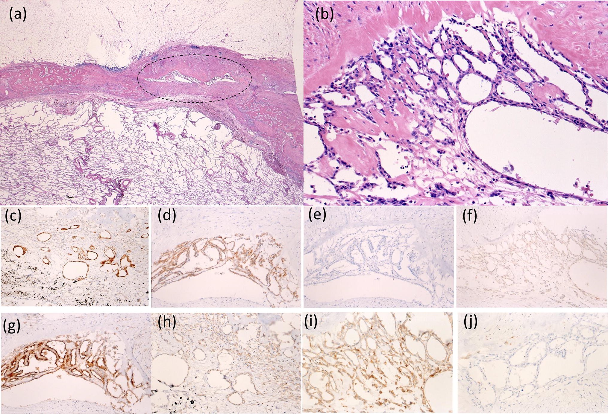
Epithelioid Mesothelioma Immunohistochemistry, Muc4 Immunohistochemistry Is Useful In Distinguishing Epithelioid Mesothelioma From Adenocarcinoma And Squamous Cell Carcinoma Of The Lung Scientific Reports
Epithelioid mesothelioma immunohistochemistry Indeed lately has been hunted by users around us, perhaps one of you. People are now accustomed to using the net in gadgets to see image and video information for inspiration, and according to the name of the post I will talk about about Epithelioid Mesothelioma Immunohistochemistry.
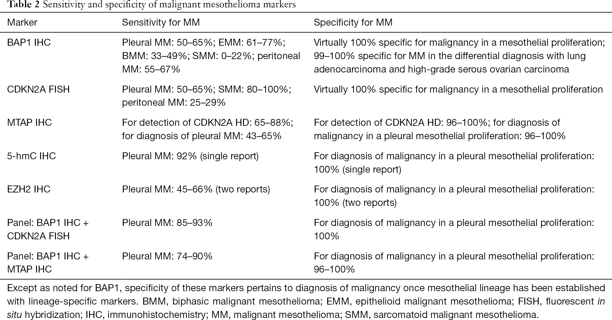
- Application Of Immunohistochemistry In Diagnosis And Management Of Malignant Mesothelioma Chapel Translational Lung Cancer Research
- Sarcomatoid Mesothelioma Get Treatment And Financial Help
- 2
- Https Academic Oup Com Ajcp Article Pdf 112 1 75 24980046 Ajcpath112 0075 Pdf
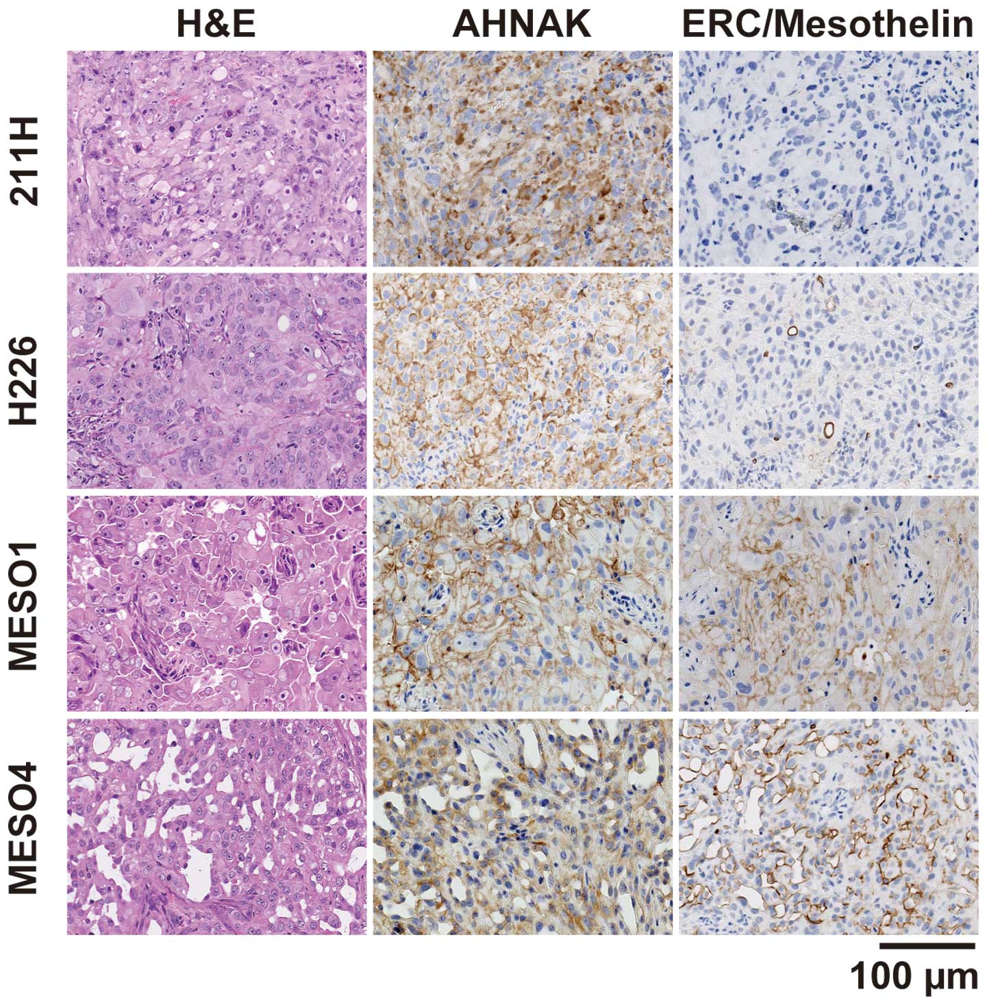
- Ahnak Is Highly Expressed And Plays A Key Role In Cell Migration And Invasion In Mesothelioma
- Zfv03is5s Mwm
Find, Read, And Discover Epithelioid Mesothelioma Immunohistochemistry, Such Us:
- What Is Epithelioid Mesothelioma Canceroz
- Mesothelioma Wikipedia
- Cient Periodique
- Muc4 Immunohistochemistry Is Useful In Distinguishing Epithelioid Mesothelioma From Adenocarcinoma And Squamous Cell Carcinoma Of The Lung Scientific Reports
- Epithelioid Mesothelioma The Most Treatable Mesothelioma
- David Meyers Md
- Simmons Beautyrest Westbrook
- Mesothelioma Develops
- Hartford Mesothelioma Attoreny
- Mesothelioma Cancer Research
If you are searching for Mesothelioma Cancer Research you've arrived at the perfect place. We ve got 104 graphics about mesothelioma cancer research including pictures, photos, pictures, backgrounds, and much more. In these webpage, we additionally provide variety of images available. Such as png, jpg, animated gifs, pic art, logo, blackandwhite, transparent, etc.

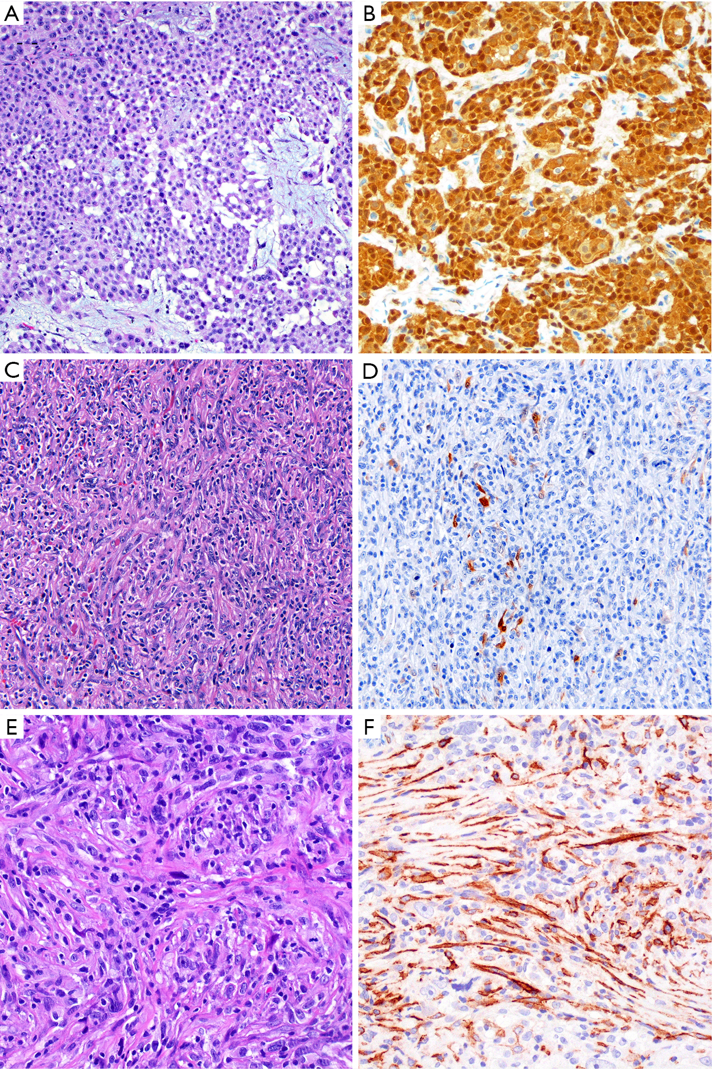
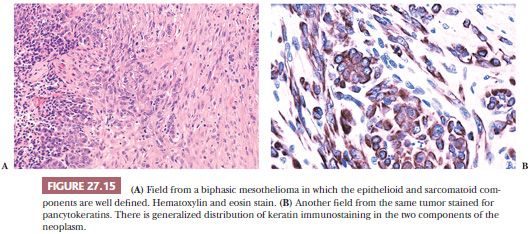
Malignant Mesothelioma And Other Mesothelial Proliferations Chapter 28 Modern Soft Tissue Pathology Mesothelioma Cancer Research
Https Academic Oup Com Ajcp Article Pdf 112 1 75 24980046 Ajcpath112 0075 Pdf Mesothelioma Cancer Research
A review and update.
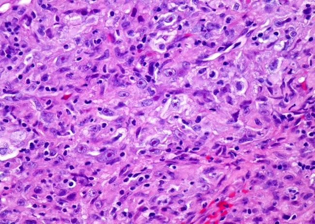
Mesothelioma cancer research. Google scholar ordonez n. J clin pathol 2002. Immunohistochemistry in the distinction between malignant mesothelioma and pulmonary adenocarcinoma.
A critical evaluation of new antibodies. The immunohistochemical diagnosis of mesothelioma. After analyzing the results it is concluded that from a practical point of view a combination of two positive mesothelioma markers wt1 and calretinin or mesothelin with two negative mesothelioma markers p63 and moc 31 would allow the differential diagnosis to be established between epithelioid mesotheliomas and squamous carcinomas of the.
After analyzing the results it is concluded that calretinin cytokeratin 56 and wt1 are the best positive markers for differentiating epithelioid malignant mesothelioma from pulmonary adenocarcinoma. Expressed in epithelioid mesothelioma but negative in sarcomatoid mesothelioma podoplanin d2 40. Keratin 5 6.
Immunohistochemistry can identify cell type and differentiate mesothelioma from other malignancies such as adenocarcinoma. Sarcomatoid cells may also be present. Membranous and apical staining.
Am j surg pathol 2003271031 51. The imig guideline has suggested the use of calretinin d2 40 wt1 and ck56 as mesothelial markers ttf 1 nap. The aims of this study were to clarify the usefulness of immunohistochemistry in the differential diagnosis of epithelioid mesothelioma with a solid growth pattern solid epithelioid mesothelioma sem and poorly differentiated squamous cell carcinoma pdscc and to confirm the validity of a specific type of antibody panel.
1 frank invasion is regarded as the most. Application of immunohistochemistry in the diagnosis of epithelioid mesothelioma. Epithelioid mesothelioma is caused by asbestos and is the most common type of the disease.
A comparative study of epithelioid mesothelioma and lung adenocarcinoma. Epithelial mesothelioma cells can develop in the lining of the lungs abdomen or heart. Of 217 cases circulated among all members of the uscanadian mesothelioma reference panel there was some disagreement about whether the process was benign or malignant in 22 of cases.
Additionally we aimed to clarify the pitfalls of. The immunohistochemical diagnosis of mesothelioma. The distinction between reactive mesothelial hyperplasia mh and malignant mesothelioma mm may be very difficult based only on histologic and morphologic findings.
The differential diagnosis of epithelioid mesothelioma from lung adenocarcinoma and squamous cell carcinoma requires the positive and negative immunohistochemical markers of mesothelioma.
More From Mesothelioma Cancer Research
- Malignant Mesothelioma Mortality
- Deadpool Coloring Pages For Adults
- Coloring Book Coloring Book
- Crayola Coloring Pages Free
- Non Squamous Cell
Incoming Search Terms:
- Epithelioid Mesothelioma Treatment Prognosis Diagnosis Non Squamous Cell,
- Guidelines For The Diagnosis And Treatment Of Malignant Pleural Mesothelioma Van Zandwijk Journal Of Thoracic Disease Non Squamous Cell,
- 2016 Evening Specialty Conference Dermatopathology Non Squamous Cell,
- Ahnak Is Highly Expressed And Plays A Key Role In Cell Migration And Invasion In Mesothelioma Non Squamous Cell,
- Utility Of Survivin Bap1 And Ki 67 Immunohistochemistry In Distinguishing Epithelioid Mesothelioma From Reactive Mesothelial Hyperplasia Non Squamous Cell,
- The Histologic And Immunohistochemical Features Of Malignant Download Scientific Diagram Non Squamous Cell,
