Ema Staining In Mesothelioma, Positive Staining Of Mesothelial Cells Malignant Mesothelioma For Download Scientific Diagram
Ema staining in mesothelioma Indeed lately is being hunted by users around us, perhaps one of you personally. People are now accustomed to using the net in gadgets to see video and image data for inspiration, and according to the title of the article I will discuss about Ema Staining In Mesothelioma.
- Pathology Outlines Epithelial Membrane Antigen Ema
- Malignant Peritoneal Mesothelioma In A Patient With Intestinal Fistula Incisional Hernia And Abdominal Infection A Case Report
- Pathology Outlines Epithelial Membrane Antigen Ema
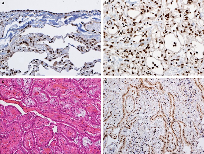
- Bap1 Brca1 Associated Protein 1 Is A Highly Specific Marker For Differentiating Mesothelioma From Reactive Mesothelial Proliferations Modern Pathology
- Different Expression Patterns Of Cd146 And Ema A D Malignant Download Scientific Diagram
- Https Pdfs Semanticscholar Org 248c 7f4791811b1339a011d6ce9b7af914c0fd40 Pdf
Find, Read, And Discover Ema Staining In Mesothelioma, Such Us:
- Positive Staining Of Mesothelial Cells Malignant Mesothelioma For Download Scientific Diagram
- Https Encrypted Tbn0 Gstatic Com Images Q Tbn 3aand9gctypj5odv93rdwye99hkagcypkwbwa4w6ydyhb1zqeu1vtgj2sd Usqp Cau
- Https Pdfs Semanticscholar Org 248c 7f4791811b1339a011d6ce9b7af914c0fd40 Pdf
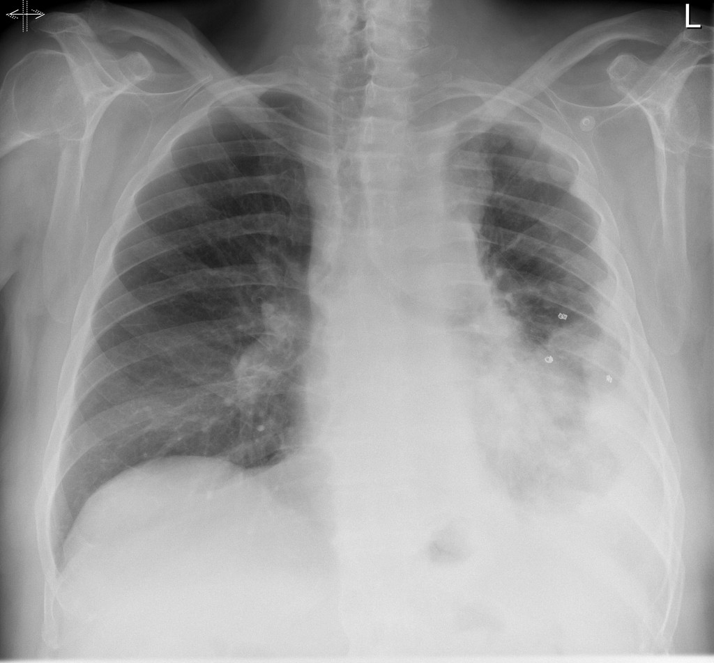
- Epithelioid Mesothelioma Radiology Case Radiopaedia Org
- Https Www Tandfonline Com Doi Pdf 10 1080 2162402x 2017 1373235
- Mandala Colouring Books For Adults
- Mesothelioma After Death
- Trademark Attorney Resume
- Color Pictures Online
- Mesothelioma Is There A Cure
If you re looking for Mesothelioma Is There A Cure you've arrived at the perfect place. We ve got 103 images about mesothelioma is there a cure adding images, photos, photographs, wallpapers, and much more. In such page, we additionally have number of graphics out there. Such as png, jpg, animated gifs, pic art, symbol, black and white, translucent, etc.

Combined Serum And Immunohistochemical Differentiation Between Reactive And Malignant Mesothelial Proliferations Sciencedirect Mesothelioma Is There A Cure
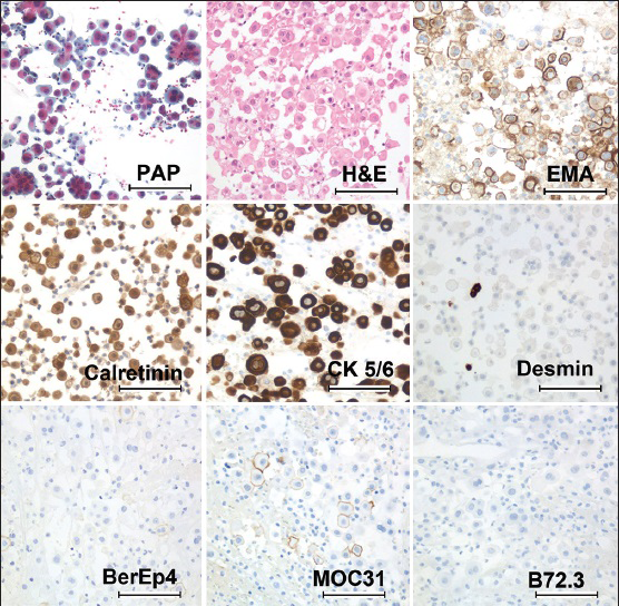
The combination of positive ema and negative desmin strongly favors mm.

Mesothelioma is there a cure. Papillary neoplasm of the peritoneum or pleura in a woman. Likewise strong membranous positivity for glut 1 andor strong nuclear staining for p53 favors a mesothelioma. Metastatic invasion ovarian neoplasm.
N corrin b nicholson a. Ema is a high molecular weight transmembranous glycosylated protein of the breast mucin complex which is useful for epithelial differentiation and has been found to be present on both carcinoma and mesothelioma cells. 73 for cd44s 73 for n cadherin 55 for vimentin 40 for e cadherin 18 for ber ep4 8 for moc 31 7 for bg 8 and none for cea b723 leu m1 ttf 1 or ca19 9.
Calretinine wt1 pos. Conversely a combination of negative ema and positive desmin favors a reactive process. Additional immunohistochemical stains should be used to differentiate mesothelial cells from carcinoma.
The diagnostic utility is limited by the wide range of tumours expressing ema and by the availability of more specific markers of epithelial differentiation. Cm cytoplasmic stain pasdiastase ema. It is concluded that strong diffuse linear staining for ema is a good marker of malignancy when differentiating epithelioid malignant mesothelioma and mesothelioma in situ from reactive mesothelial hyperplasia although weak focal staining may occur in reactive conditions.
B i o p s y. To our knowledge this case of peritoneal mesothelioma is the first to describe an opposite immunohistochemical profile to that expected being ema negative and desmin positive despite macroscopic and microscopy evidence of malignancy. Ema staining in mesothelioma.
The use of epithelial membrane antigen and silver stained nucleolar organizer regions testing in the differential diagnosis of mesothelioma from benign reactive mesothelioses. A microcystic meningioma showing positive cytoplasmic and membranous staining for ema 200 b transitional meningioma featuring cytoplasmic reactivity for d2 40 200 c and d meningothelial cells showing focal cytoplasmic staining for keratin 56 and mesothelioma ab1 respectively 100. Immunohistochemical expression of ema and mesothelioma related markers in meningioma cases.
The membranous staining is said to be much stronger in mesothelioma 4. All 100 of the mesotheliomas reacted for calretinin cytokeratin 56 and mesothelin 93 for wt1 93 for ema 85 for hbme 1 77 for thrombomodulin.

Guidelines For Cytopathologic Diagnosis Of Epithelioid And Mixed Type Malignant Mesothelioma Complementary Statement From The International Mesothelioma Interest Group Also Endorsed By The International Academy Of Cytology And The Papanicolaou Society Of Mesothelioma Is There A Cure
More From Mesothelioma Is There A Cure
- Mags Portman Mesothelioma Blog
- Flower Garden Coloring Pages
- Dog Coloring Pages Of Animals
- Tips For Mesothelioma Caregivers
- Food Coloring Pages For Adults
Incoming Search Terms:
- Immunohistochemical Staining For Muc1 Ema On Sections Of Formalin Fixed Download Scientific Diagram Food Coloring Pages For Adults,
- Application Of Immunohistochemistry In Diagnosis And Management Of Malignant Mesothelioma Chapel Translational Lung Cancer Research Food Coloring Pages For Adults,
- Https Www Karger Com Article Pdf 377697 Food Coloring Pages For Adults,
- Immunohistochemical Evaluation For Mpm Vs Reactive Mesothelium Download Scientific Diagram Food Coloring Pages For Adults,
- Pleomorphic And Desmoplastic Malignant Mesotheliomas And A Malignant Mesothelioma With Osseous And Cartilaginous Differentiation Case Reports Food Coloring Pages For Adults,
- Expression Of Mesothelioma Related Markers In Meningiomas An Immunohistochemical Study Food Coloring Pages For Adults,


