Ct Images Of Mesothelioma, Https Encrypted Tbn0 Gstatic Com Images Q Tbn 3aand9gcs1t0mw4md7 2uqg1nzj6xmvh3xaabyt Pap8 X 68 Usqp Cau
Ct images of mesothelioma Indeed recently is being hunted by consumers around us, maybe one of you. Individuals are now accustomed to using the net in gadgets to view image and video data for inspiration, and according to the name of the article I will discuss about Ct Images Of Mesothelioma.
- Pleural Effusion In Mesothelioma Ct Scan Stock Image C013 9670 Science Photo Library
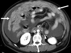
- Primary Peritoneal Mesothelioma Radiology Case Radiopaedia Org
- Malignant Pleural Mesothelioma Evaluation With Ct Mr Imaging And Pet Radiographics
- Diagnostic Imaging And Workup Of Malignant Pleural Mesothelioma
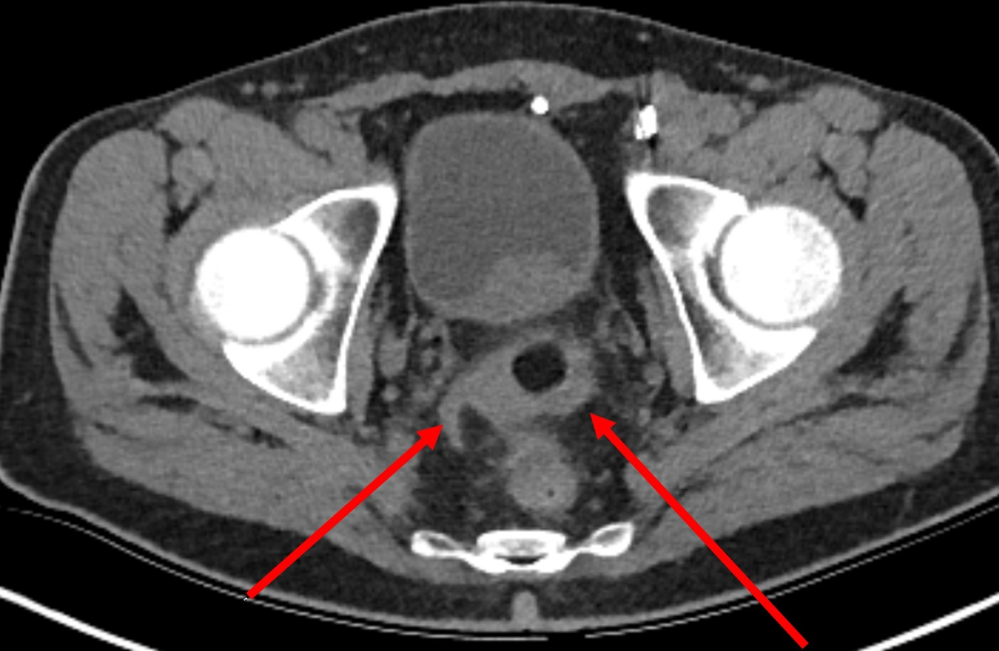
- Cureus Prostate Carcinoma And Pleural Mesothelioma An Extremely Rare Co Occurrence
- Assessment Of Therapy Responses And Prediction Of Survival In Malignant Pleural Mesothelioma Through Computer Aided Volumetric Measurement On Computed Tomography Scans Journal Of Thoracic Oncology
Find, Read, And Discover Ct Images Of Mesothelioma, Such Us:
- Chest Ct Scan Mesothelioma Due To Asbestos Atb578 Rayandmave S Blog
- Mesothelioma With Radiation Hepatitis Changes In The Liver Chest Case Studies Ctisus Ct Scanning
- Malignant Mesothelioma Radiographics
- Pleural Effusion In Mesothelioma Ct Scan Stock Image C013 9667 Science Photo Library
- Malignant Pleural Mesothelioma Evaluation With Ct Mr Imaging And Pet Radiographics
- Christmas Ornament Coloring Pages
- Happy Hanukkah Coloring Pages
- Online Coloring Book For Kids
- Tecentriq Sclc
- Isaacs And Isaacs Law Firm
If you re looking for Isaacs And Isaacs Law Firm you've come to the ideal location. We ve got 104 graphics about isaacs and isaacs law firm including pictures, pictures, photos, wallpapers, and more. In these webpage, we also have variety of graphics out there. Such as png, jpg, animated gifs, pic art, symbol, black and white, translucent, etc.
Mesothelioma also known as malignant mesothelioma is an aggressive malignant tumor of the mesothelium.

Isaacs and isaacs law firm. 12 clinical characteristics treatment and survival outcomes in malignant mesothelioma. There are five primary types of imaging tests used during a mesothelioma diagnosis including x rays mri scans ct scans pet scans and ultrasoundsimaging scans are non invasive and act as a stepping stone in the diagnostic process for mesothelioma patients. Ct scans are more than 90 sensitive for detecting malignant pleural mesothelioma.
X rays also indicate fluid build up another type of asbestos related condition which may indicate mesothelioma. Development of a guideline on reading ct images of malignant pleural mesothelioma and selection of the reference ct films european journal of radiology vol. However appearances can be subjective and are highly operator dependent he said.
The 3 d images ct scans provide a far more detailed view and offer more than 90 percent detection sensitivity. Pleural mesothelioma 90 covered in this article. Given the presence of the mesothelium in different parts of the body mesothelioma can arise in various locations 17.
The images are used to provide a better understanding of mesothelioma cancer how it develops and treatment options as well as imagery around asbestos and other related topics. The x ray images are put into a computer that takes all the images and makes a 3d image of your chest. Most tumors arise from the pleura and so this article will focus on pleural mesothelioma.
Below is a collection of images related to mesothelioma and asbestos for reference and convenience. This image details the anatomy of the lung and specifically highlights the lining of the lungs known as the pleura where pleural mesothelioma developsthis is the most common form of the cancer and develops when the mesothelial cells that line the pleura become cancerous and divide uncontrollably. Furthermore although less effective for detecting peritoneal mesothelioma ct scans are still the most useful imaging study for diagnosing malignant.
The 3d image obtained from a ct scan provides a far more detailed view of a persons internal anatomy than a simple two dimensional x ray image. Some ct scans may use various contrast materials to help the device tell the difference between organs and body structures. This material is able to differentiate between small mesothelioma tumors and body organs better.
Types of mesothelioma imaging tests. Computed tomography or ct scans also uses x rays. Eighteen years experience in turkey.
Diagnostic ct scan for mesothelioma.

Localized Malignant Pleural Sarcomatoid Mesothelioma Misdiagnosed As Benign Localized Fibrous Tumor Kim Journal Of Thoracic Disease Isaacs And Isaacs Law Firm
More From Isaacs And Isaacs Law Firm
- Richards Law
- Mesothelioma Abdominal Cancer
- Junior Lawyer Lawyer Cv Sample Pdf
- Mesothelioma Cancer Doctors
- Cost To Remove Vermiculite
Incoming Search Terms:
- Malignant Pleural Mesothelioma Radiology Case Radiopaedia Org Cost To Remove Vermiculite,
- Volumetry An Alternative To Assess Therapy Response For Malignant Pleural Mesothelioma European Respiratory Society Cost To Remove Vermiculite,
- Localized Malignant Mesothelioma An Unusual And Poorly Characterized Neoplasm Of Serosal Origin Best Current Evidence From The Literature And The International Mesothelioma Panel Modern Pathology Cost To Remove Vermiculite,
- The Role Of Imaging In Malignant Pleural Mesothelioma An Update After The 2018 Bts Guidelines Clinical Radiology Cost To Remove Vermiculite,
- Learningradiology Mesothelioma Radiology Malignant Imaging X Ray Asbestosis Cost To Remove Vermiculite,
- A 71 Year Old Patient With Pleural Mesothelioma Ct Scan Top A Download Scientific Diagram Cost To Remove Vermiculite,


